. 2004 Dec 13;91(12):2086-93.
doi: 10.1038/sj.bjc.6602232.

MUC1 expression in primary and metastatic pancreatic cancer cells for in vitro treatment by (213)Bi-C595 radioimmunoconjugate

Affiliations

MUC1 expression in primary and metastatic pancreatic cancer cells for in vitro treatment by (213)Bi-C595 radioimmunoconjugate

C F Qu et al. Br J Cancer. .

Abstract

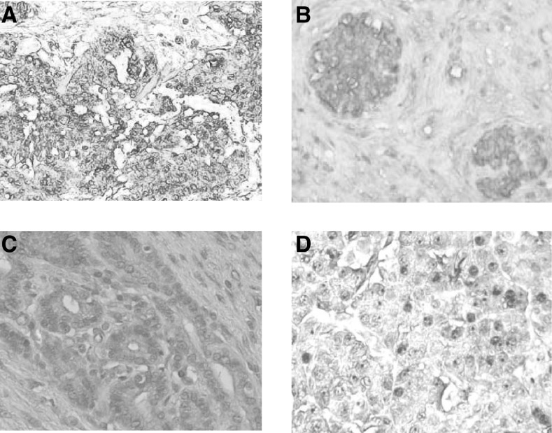
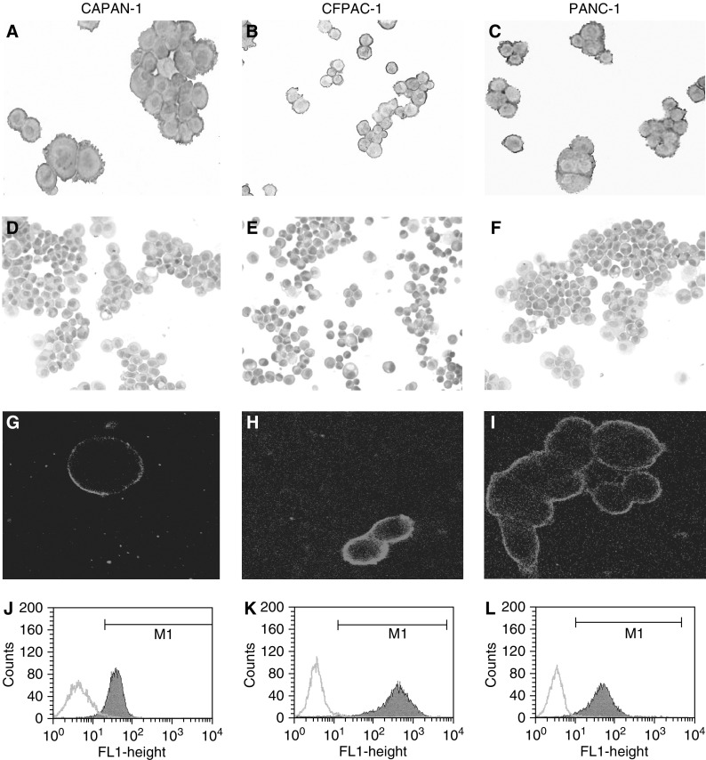
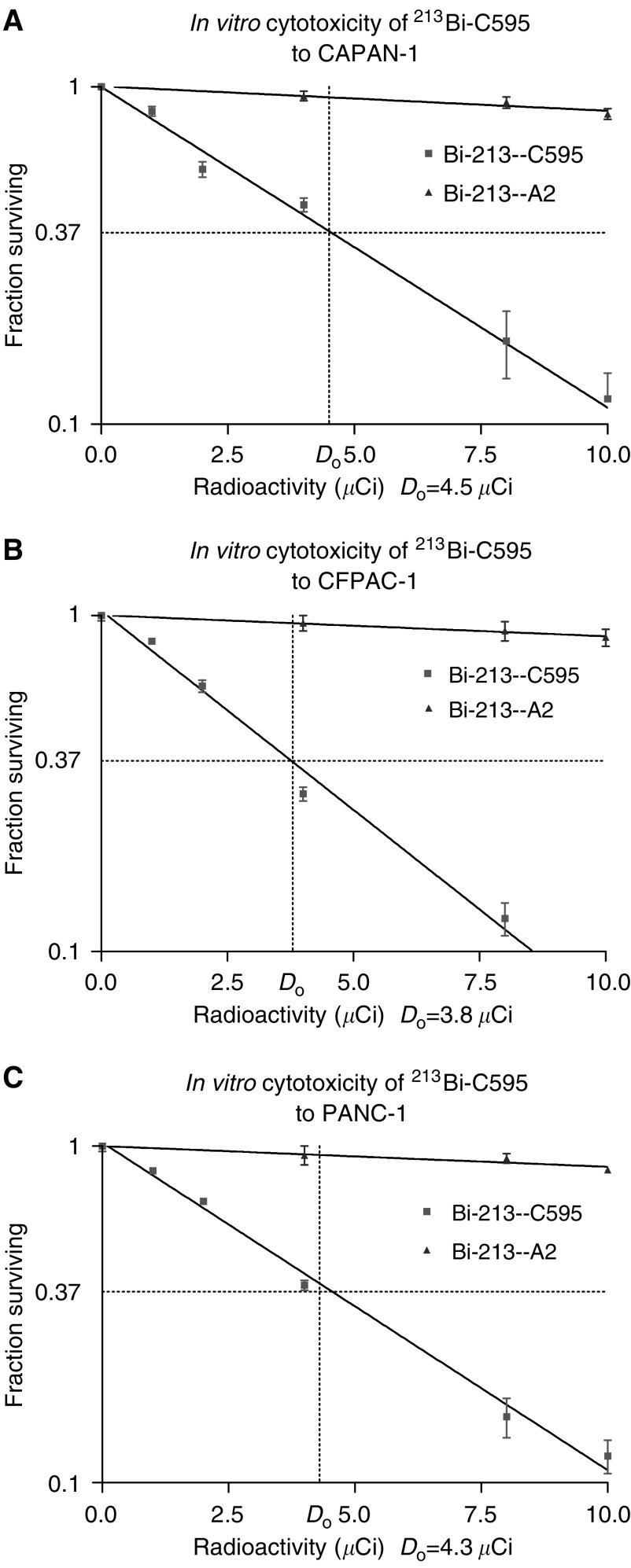
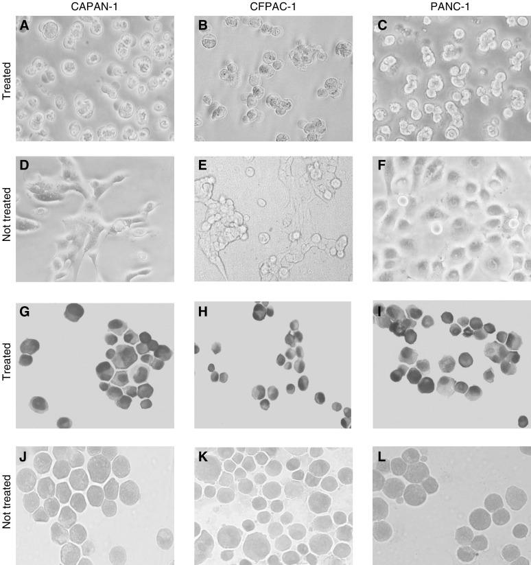
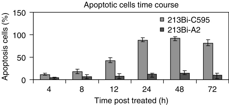
Control of micrometastatic pancreatic cancer remains a major objective in pancreatic cancer treatment. The overexpression of MUC1 mucin plays an important role in cancer metastasis. The aim of this study was to detect the expression of MUC1 in human primary tumour tissues and three pancreatic cancer cell lines (CAPAN-1, CFPAC-1 and PANC-1), and target MUC1-positive cancer cells in vitro using (213)Bi-C595 alpha-immunoconjugate (AIC). The expression of MUC1 on pancreatic tumour tissues and cancer cell lines was performed by immunohistochemistry and further confirmed by confocal microscope and flow cytometry analysis on the cell surface. Cytotoxicity of (213)Bi-C595 was tested by MTS assay. Apoptosis was documented using TUNEL assay. Overexpression of MUC1 was found in approximately 90% of tested tumour samples and the three pancreatic cancer cell lines. (213)Bi-C595 is specifically cytotoxic to pancreatic cancer cells in a concentration-dependent fashion. These results suggest that overexpression of MUC1 in pancreatic cancer is a useful target, and that the novel (213)Bi-C595 AIC selectively targets pancreatic cancer cells in vitro. (213)Bi-C595 may be a useful agent for the treatment of micrometastases or minimal residual disease (MRD) in pancreatic cancer patients with overexpression of MUC1 antigen.


Figures

Figure 1
Figure 1
Expression of MUC1 in pancreatic cancer tissues. MUC1 expression was assessed by immunohistochemistry. Representative pictures are shown for immmunostaining with MAbs C595 (test) and A2 (control). High expression of MUC1 was found in patient 1 staining with MAb C595 (A) and moderate expression was found in patient 2 staining with MAb C595 (B), while no MUC1 expression was found in patient staining with MAb A2 (C) or in normal pancreas (D).
Figure 2
Figure 2
Expression of MUC1 in pancreatic cancer cell lines. Representative pictures are shown for immmunostaining with MAbs C595 (test) and A2 (control). MAb C595 was strongly positive in three cell lines (A–C), while MAb A2 was negative in three cancer cell lines (D–F). Brown staining indicates positive cells. The MUC1 cell surface expression in three cancer cell lines was confirmed by Confocal Microscope (G–I). Green staining indicates positive cells. Expression of MUC1 in three viable pancreatic cancer cell lines was assessed by FACA analysis (J–L). Data are presented as histograms, using a mouse IgG1-negative control to determine background fluorescence and to set the marker (M1). All of the photographs are at × 200 magnification.
Figure 3
Figure 3
Representative cytotoxicity study of CAPAN-1 (A), CFPAC-1 (B) and PANC-1 (C) cells following treatment for 24 h with 213Bi-C595. Cells were treated with varying concentrations of 213Bi-C595 or nonspecific control 213Bi-A2, incubated overnight and cell survival was measured by MTS assay at 24 h and expressed as a percentage of cell survival of control cells. Results are expressed as a mean percent s.d. of control plates containing nonspecific α conjugates. Each experiment was performed in triplicate, and each point represents the mean of three experiments.
Figure 4
Figure 4
Morphological changes in three cell lines incubated with 213Bi-C595 (A–C) for 24 hours. (D–F) Matched, untreated cells are shown. TUNEL assays of CAPAN-1, CFPAC-1 and PANC-1 cells are shown in (G–I), and control in (J–L). Typical apoptotic cells with condensed or fragmented nuclei are observed in treated cell lines, while control cells show normal shapes. All of the photographs are at × 200 magnification.
Figure 5
Figure 5
Observation of apoptosis in time course after treatment with 213Bi-C595 and 213Bi-A2 AICs. Blue and red columns represent 213Bi-C595 and 213Bi-A2, respectively. The PANC-1 cells were treated with 213Bi-C595 and 213Bi-A2 with concentrations of 10 μCi 105cells−1, incubated at 4, 8, 12, 24, 48 and 72 h, tested by TUNEL assay, and expressed as a percentage of TUNEL-positive cells in total cells. Each experiment was performed in triplicate, and each point represents the mean of three experiments.

References

    1. Abbas Rizvi SM, Sarkar S, Goozee G, Allen BJ (2000) Radioimmunoconjugates for targeted alpha therapy of malignant melanoma. Melanoma Res 10: 281–289 - PubMed
    1. Allen BJ (1999) Can alpha-immunotherapy succeed where other systemic modalities have failed? Nucl Med Commun 20: 205–207 - PubMed
    1. Boll RA, Mirzaden S, Kennel SJ (1997) Optimizations of radiolabeling of immunoproteins with 213Bi. Radiochimica Acta 79: 145–149
    1. Farah RA, Clinchy B, Herrera L, Vitetta ES (1998) The development of monoclonal antibodies for the therapy of cancer. Crit Rev Eukar Gene Expr 8: 321–356 - PubMed
    1. Feng H, Ghazizadeh M, Konishi H, Araki T (2002) Expression of MUC1 and MUC2 mucin gene products in human ovarian carcinomas. Jpn J Clin Oncol 32: 525–529 - PubMed

Publication types