. 2004 Dec;114(12):1714-25.
doi: 10.1172/JCI22123.

Platelet-derived lysophosphatidic acid supports the progression of osteolytic bone metastases in breast cancer

Affiliations

Platelet-derived lysophosphatidic acid supports the progression of osteolytic bone metastases in breast cancer

Ahmed Boucharaba et al. J Clin Invest. 2004 Dec.

Abstract

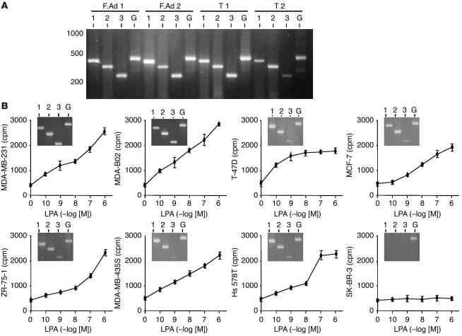
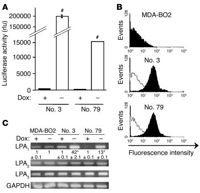
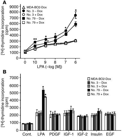
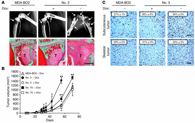
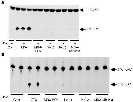
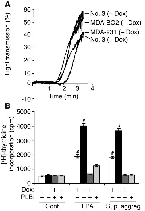
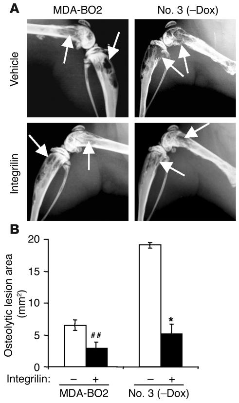
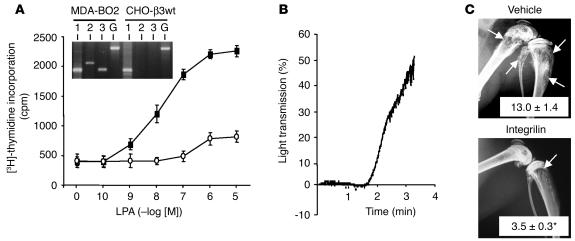
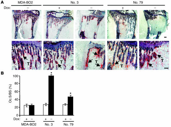
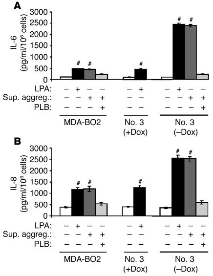
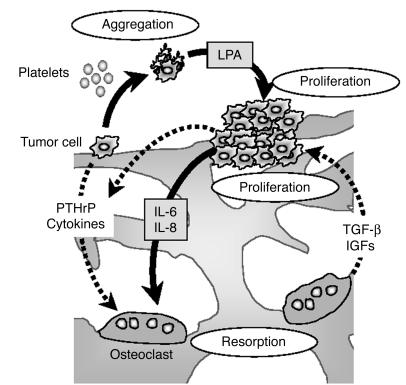
The role of lysophosphatidic acid (LPA) in cancer is poorly understood. Here we provide evidence for a role of LPA in the progression of breast cancer bone metastases. LPA receptors LPA(1), LPA(2), and LPA(3) were expressed in human primary breast tumors and a series of human breast cancer cell lines. The inducible overexpression of LPA(1) in MDA-BO2 breast cancer cells specifically sensitized these cells to the mitogenic action of LPA in vitro. In vivo, LPA(1) overexpression in MDA-BO2 cells enhanced the growth of subcutaneous tumor xenografts and promoted bone metastasis formation in mice by increasing both skeletal tumor growth and bone destruction. This suggested that endogenous LPA was produced in the tumor microenvironment. However, MDA-BO2 cells or transfectants did not produce LPA. Instead, they induced the release of LPA from activated platelets which, in turn, promoted tumor cell proliferation and the LPA(1)-dependent secretion of IL-6 and IL-8, 2 potent bone resorption stimulators. Moreover, platelet-derived LPA deprivation in mice, achieved by treatment with the platelet antagonist Integrilin, inhibited the progression of bone metastases caused by parental and LPA(1)-overexpressing MDA-BO2 cells and reduced the progression of osteolytic lesions in mice bearing CHO-beta3wt ovarian cancer cells. Overall, our data suggest that, at the bone metastatic site, tumor cells stimulate the production of LPA from activated platelets, which enhances both tumor growth and cytokine-mediated bone destruction.


Figures

Figure 1
Figure 1
Expression of LPA receptors in breast cancer and mitogenic activity of LPA in breast cancer cell lines. (A) RT-PCR experiments using total RNA isolated from human primary tumors: fibroadenomas (F.Ad 1, F.Ad 2), ductal carcinomas (T 1, T 2). Expected size of amplification products for LPA1 (1), LPA2 (2), LPA3 (3), and GAPDH (G) are 428, 352, 256, and 470 bp, respectively. (B) Human breast cancer cell lines were stimulated with increasing concentrations of LPA and pulsed with [3H]-thymidine. Cell proliferation was assessed after quantification of [3H]-thymidine incorporation. Data are expressed in cpm as the mean ± SD of 6 replicates and are representative of at least 3 separate experiments. Insets: RT-PCR amplification products for LPA1, LPA2, LPA3, and GAPDH using total RNA isolated from each indicated cell line.
Figure 2
Figure 2
Characterization of MDA-BO2 clones stably transfected to conditionally overexpress HA-LPA1. (A) Cells transfected with the bidirectional expression vector pBiL-HA-LPA1 were plated with (+) or without (–) doxycycline (Dox). Two stable clones (nos. 3 and 79) were selected using luciferase activity measurement as an end point. Data are expressed in relative light units (rlu). #P < 0.0001 for cells without doxycycline versus cells with doxycycline. (B) Detection of HA-LPA1 cell surface expression in parental MDA-BO2 cells and in clones no. 3 and no. 79 by flow cytometry using the anti-HA monoclonal antibody. Black and white histograms refer to cells treated without and with doxycycline, respectively. The y axis depicts the number of cells per channel (events), and the x axis depicts the relative fluorescence intensity in arbitrary units (log scale). (C) LPA receptor mRNA expression in parental MDA-BO2 cells and in clones no. 3 and no. 79. Cells were cultured in the absence or presence of doxycycline before total RNA preparation. RT-PCR fragments were separated on a 2% agarose gel and then stained with ethidium bromide. Numbers below the top panel correspond to real-time PCR quantification data of the LPA1 mRNA copy number for each clone compared with that of the parental MDA-BO2 cells cultured in the absence of doxycycline (mean ± SD; *P < 0.001). No variation of mRNA expression was detected for LPA2, LPA3, or GAPDH in the presence or absence of doxycycline.
Figure 3
Figure 3
Effect of LPA1 overexpression on the mitogenic action of LPA on MDA-BO2 cells. (A) Parental MDA-BO2 cells (triangles) and transfected clones no. 3 (circles) and no. 79 (squares) were cultured in plain medium (open symbols) or medium supplemented with 100 ng/ml of doxycycline (filled symbols) and then treated as described in Figure 1B. Data are expressed in cpm as the mean ± SD of 6 replicates and are representative of at least 3 separate experiments. **P < 0.005; *P < 0.001 untreated versus doxycycline-treated cell lines. (B) Cells were cultured in the absence or presence of doxycycline (100 ng/ml) and stimulated with LPA (0.1 μM) or other indicated growth factors (10 ng/ml). Cell proliferation was measured as described above. Data are expressed as the mean ± SD of 6 replicates and are representative of 3 separate experiments. *P < 0.001 untreated versus doxycycline-treated cell lines. Cont., control.
Figure 4
Figure 4
Effect of LPA1 overexpression in MDA-BO2 cells on osteolytic lesions and on skeletal and subcutaneous tumor growth. (A) Animals fed without or with doxycycline were inoculated intravenously with MDA-BO2 or clone no. 3 cells. (Upper panels) Representative radiographs of hind limbs from mice bearing MDA-BO2 or clone no. 3 cells 30 days after tumor cell inoculation. In the absence of doxycycline, there was a marked increase in the extent of osteolytic lesions (arrows) in mice bearing clone no. 3 cells. (Lower panels) Representative bone histology of Goldner’s trichrome–stained tibial metaphysis from metastatic animals. Bone is stained in green; bone marrow and tumor cells are stained red. Trabecular bone was completely destroyed and replaced by tumor cells (T) in tibial metaphysis from untreated animals bearing clone no. 3 cells. Scale bar: 1 mm. (B) Animals fed without (open symbols) or with (filled symbols) doxycycline were inoculated subcutaneously into the flank with MDA-BO2 (triangles), clone no. 3 (circles), or clone no. 79 (squares) cells. Tumors were measured at the indicated time points. V (in mm3) is expressed as the mean ± SD of 9 animals per group. P < 0.05; **P < 0.005 doxycycline-free versus doxycycline-fed tumor-bearing animals. (C) Subcutaneous (upper panels) and skeletal (lower panels) tumor tissue sections immunostained with an antibody against the nuclear Ki-67 antigen. The mitotic index (numbers on each panel) was calculated as the percentage of nuclei positive for Ki-67. Results are the mean ± SD of 6 independent tumor sections. #P < 0.0001 doxycycline-free versus doxycycline-fed tumor-bearing animals. Scale bars: 100 μm.
Figure 5
Figure 5
Production of LPA and ATX by MDA-BO2 breast cancer cells. Cell culture media were collected for each indicated cell line placed in the absence or presence of doxycycline (100 ng/ml). (A) Detection of LPA. The production of phosphatidic acid (PA) was due to the transfer of [14C] fatty acyl chain (FA) onto LPA present in the reaction mixture assay. Experiments were carried out in duplicate for each cell line. Purified LPA (50, 100, and 200 pmol) was used as a positive control. DMEM (Cont.) was used as a negative control. (B) Measurement of ATX/lyso-PLD activity. [14C]-LPC was used as the substrate of ATX/lyso-PLD to produce [14C]-LPA. Experiments were carried out in duplicate using culture media from the cell lines described above. Note that MDA-MB-231, MDA-BO2, clone no. 3, and clone no. 79 cell lines did not produce LPA or express ATX.
Figure 6
Figure 6
Effect of breast tumor cells on platelet aggregation and the release of LPA from activated platelets. (A) Indicated tumor cells previously cultured in the absence or presence of doxycycline were added to washed human platelets under stirring conditions. Platelet aggregation was recorded over the time as the percentage of light transmission. (B) Clone no. 3 cells were plated without or with doxycycline and stimulated with DMEM, LPA (10–7 M) or MDA-BO2–induced platelet aggregation supernatants (Sup. aggreg.), in the presence or absence of PLB. Cell proliferation was measured as described in the legend of Figure 3. Data are expressed as the mean ± SD of 6 replicates and are representative of 3 separate experiments. #P < 0.0001, stimulated versus control cells.
Figure 7
Figure 7
Effect of in vivo inhibition of platelet aggregation on the LPA-dependent progression of breast cancer bone metastases. (A) Representative radiographs at day 30 of hind limbs from doxycycline-free fed mice bearing MDA-BO2 or clone no. 3 cells that were treated with Integrilin or vehicle from day 14 to day 30. (B) Quantification of osteolytic lesion areas on radiographs in Integrilin-treated (+) and vehicle-treated (–) metastatic animals. Values are the mean ± SE of 6–9 animals per group. ##P < 0.01; *P < 0.001, Integrilin-treated versus vehicle-treated animals.
Figure 8
Figure 8
Mitogenic effect of LPA on CHO-β3wt cells in vitro and effect of in vivo inhibition of platelet aggregation on the progression of CHO-β3wt bone metastases. (A) Cell proliferation assay: CHO-β3wt cells were incubated with increasing concentrations of LPA in the absence (filled squares) or presence (open circles) of PLB. Cell proliferation was assessed as described in Figure 1B. Data are expressed in cpm as the mean ± SD of 6 replicates and are representative of at least 3 separate experiments. Inset: RT-PCR amplification products for LPA1, LPA2, LPA3, and GAPDH using MDA-BO2 or CHO-β3wt total RNAs. Expected size of amplification products for LPA1, LPA2, LPA3, and GAPDH are 192, 282, 182, and 470 bp, respectively. (B) CHO-β3wt cell stimulation of platelet aggregation was carried out as described in Figure 6. (C) Bone metastasis experiment. Representative radiographs at day 21 of hind limbs from mice bearing CHO-β3wt cells treated with Integrilin or vehicle from day 10 to day 21. Osteolytic lesions are indicated by arrows. Data represent the mean ± SE of osteolytic lesion areas, expressed in mm2, of 8–10 animals per group. *P < 0.001 Integrilin-treated versus vehicle-treated animals.
Figure 9
Figure 9
Effect of LPA1 overexpression in MDA-BO2 cells on osteoclast activity in vivo. (A) Animals fed without or with doxycycline were inoculated intravenously with MDA-BO2 or transfectants (clone no. 3 and clone no. 79 cells). Representative histological examination of TRAP-stained proximal tibia section from metastatic animals 30 days after tumor cell inoculation. Lower panels show magnified areas (white squares) from upper panels. Bone is stained in dark blue and osteoclasts are stained in red (arrows). Scale bar: 200 μm. (B) The Oc.S/BS ratio was quantified. Results are the mean ± SD of 4–6 animals per group. *P < 0.001 doxycycline-free versus doxycycline-fed animals.
Figure 10
Figure 10
Effect of purified or platelet-derived LPA on the production of IL-6 and IL-8 by breast cancer cells. IL-6 (A) and IL-8 (B) were quantified using culture media from cells pretreated in the presence or absence of LPA, the supernatant of breast cancer cell–induced platelet aggregation, and PLB. Data are expressed as the mean ± SD of 3 replicates and are representative of 2 separate experiments. #P < 0.0001 stimulated versus unstimulated cells.
Figure 11
Figure 11
Schematic representation of the LPA effects on progression of osteolytic bone metastases. Breast cancer cells produce factors (PTHrP, cytokines) that stimulate osteoclast-mediated bone resorption. In turn, bone resorption releases growth factors (IGFs, TGF-β) from the bone matrix that stimulate tumor growth and the production of PTHrP by tumor cells (16). This results in a vicious cycle, illustrated by dotted arrows. Bone-residing breast cancer cells also induce platelet aggregation and the release of LPA from activated platelets. Platelet-derived LPA then stimulates both tumor growth and the production of IL-6 and IL-8 by tumor cells (black arrows), which in turn enhance bone resorption.

Comment in

References

    1. An S, Dickens MA, Bleu T, Hallmark OG, Goetzl EJ. Molecular cloning of the human Edg2 protein and its identification as a functional cellular receptor for lysophosphatidic acid. Biochem. Biophys. Res. Commun. 1997;231:619–622. - PubMed
    1. An S, Bleu T, Hallmark OG, Goetzl EJ. Characterization of a novel subtype of human G protein-coupled receptor for lysophosphatidic acid. J. Biol. Chem. 1998;273:7906–7910. - PubMed
    1. Bandoh K, et al. Molecular cloning and characterization of a novel human G-protein-coupled receptor, EDG7, for lysophosphatidic acid. J. Biol. Chem. 1999;274:27776–27785. - PubMed
    1. Noguchi K, Ishii S, Shimizu T. Identification of p2y9/GPR23 as a Novel G Protein-coupled receptor for lysophosphatidic acid, structurally distant from the Edg family. J. Biol. Chem. 2003;278:25600–25606. - PubMed
    1. McIntyre TM, et al. Identification of an intracellular receptor for lysophosphatidic acid (LPA): LPA is a transcellular PPARgamma agonist. Proc. Natl. Acad. Sci. U. S. A. 2003;100:131–136. - PMC - PubMed

Publication types

MeSH terms