. 2004 Dec;114(12):1774-81.
doi: 10.1172/JCI22284.

Adoptive immunotherapy of prostate cancer bone lesions using redirected effector lymphocytes

Affiliations

Adoptive immunotherapy of prostate cancer bone lesions using redirected effector lymphocytes

Jehonathan H Pinthus et al. J Clin Invest. 2004 Dec.

Abstract

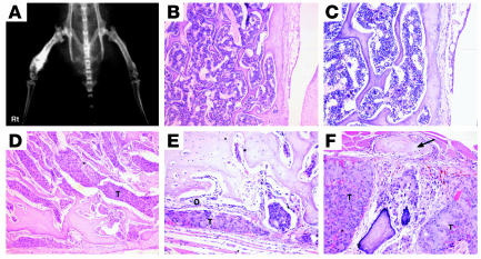
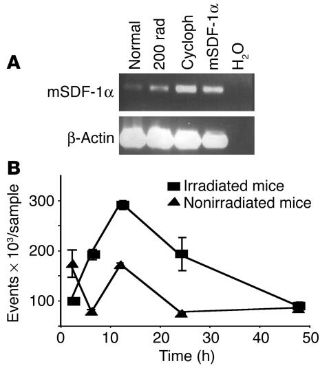
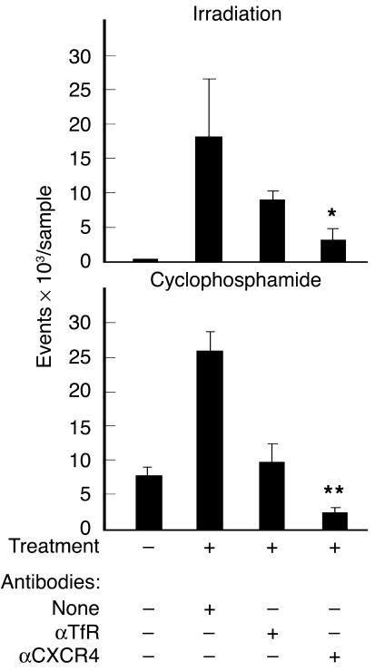
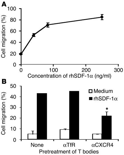
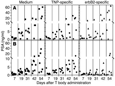
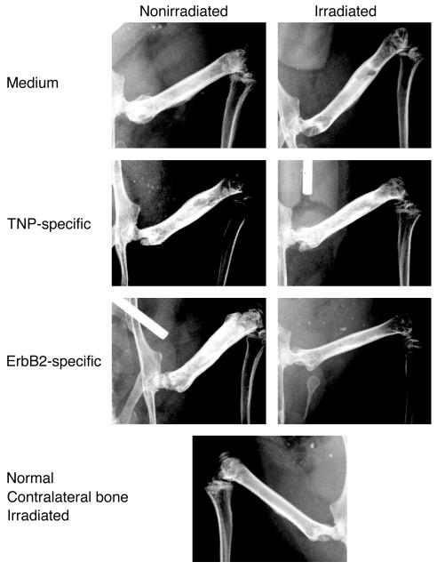
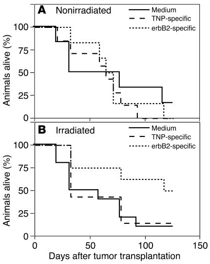
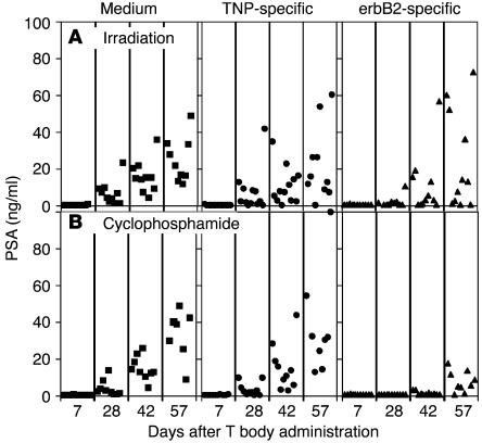
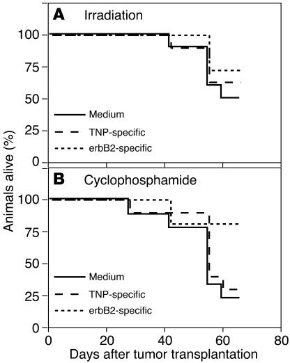
Prostate cancer is currently the most commonly diagnosed noncutaneous malignancy in American men. When metastatic, usually to the bone, the disease is no longer curable and is usually treated palliatively with androgen ablation. However, after conversion to androgen-independent disease, there is no effective therapy currently available. The "T body" approach, which uses genetically reprogrammed lymphocytes derived from the patient and expressing chimeric receptor genes, combines the effector functions of T lymphocytes and NK cells with the ability of antibodies to recognize predefined surface antigens with high specificity and in a non-MHC-restricted manner. We show here the therapeutic efficacy of human lymphocytes bearing erbB2-specific chimeric receptors on human prostate cancer BM lesions in a SCID mouse model after conditioning of the recipient to allow homing and persistent functioning of the adoptively transferred cells. Induction of stromal cell-derived factor-1 production within the BM using low-dose irradiation or cyclophosphamide combined with IL-2 administration enhanced the homing of systemically delivered T bodies, resulting in decreased tumor growth and prostate-specific antigen secretion, prolongation of survival, and even cure of the treated mice. These preclinical studies strongly support the idea that the T body approach has therapeutic potential in disseminated prostate cancer.


Figures

Figure 1
Figure 1
WISH-PC14 induces osteoblastic bone lesions. A single-cell suspension of WISH-PC14 was injected transtrochanterically into the right femur of a SCID mouse. (A) Radiographic appearance of the tumor. (B) Normal BM histology of the contralateral femur. Magnification, ×100. (C) Same as B. Magnification, ×200. Note the normal bone trabeculi and periosteum. (D) WISH-PC14 (T) occupies the entire BM cavity of the right femur. Magnification, ×100. (E) Tumor cells surrounded by osteoblastic activity (O). Magnification, ×200. Note the thickening of the bone trabeculi. (F) Same preparation and magnification as in E. Note the periosteal reaction of paraosteal bone formation (arrow).
Figure 2
Figure 2
Effect of irradiation and cyclophosphamide on SDF-1 mRNA expression in BM and subsequent migration of human T bodies to murine BM. (A and B) SCID mice were irradiated with 2 Gy or injected i.p. with 200 mg/kg of cyclophosphamide or were left untreated. (A) Twenty-four hours after the treatments, mRNA was prepared and subjected to RT-PCR. Cycloph, cyclophosphamide; mSDF-1α, mouse SDF-1α; normal, untreated mouse. H2O was used as a negative control. (B) Kinetics of the migration of human T bodies to the BM after their i.v. injection into treated mice. The number of T bodies reaching the BM (events) was determined by FACS analysis gating on GFP (n = 3 mice per group; experiment was repeated at least twice). The number of T bodies that accumulated in the BM is significantly larger in irradiated than in nonirradiated recipients at 6 and 12 hours (P = 0.054 and P = 0.011, respectively).
Figure 3
Figure 3
Migration of T bodies to the BM of preconditioned mice is mediated in part by CXCR4. SCID mice (n = 3 mice per group) were irradiated with 2 Gy TBI or were injected with 200 mg/kg cyclophosphamide, 24 hours before i.v. administration of erbB2-specific CR–bearing lymphocytes. T bodies were preincubated for 30 minutes on ice with mAb against CXCR4 (αCXCR4), irrelevant control mAb against human TfR (αTfR), or no antibody (none) and then were injected systemically into the mice. After 24 hours, BM was extracted and the number of human lymphocytes was measured by FACS analysis (detecting GFP-positive T bodies in the irradiation experiment and CFSE-labeled cells in the cyclophosphamide experiment). Treatment with anti-CXCR4 was significantly different from control treatment (anti-TfR) for both irradiated mice (*P = 0.003) and cyclophosphamide-treated mice (**P = 0.009) and was significantly different from no antibody treatment (P = 0.01 and P = 0.0002, respectively).
Figure 4
Figure 4
Transwell migration of T bodies toward recombinant human SDF-1α (rhSDF-1α) is dose dependent and is inhibited by anti-CXCR4. (A and B) Transwell migration of T bodies. (A) T bodies migrate toward increasing concentrations of rhSDF-1α. (B) T bodies were preincubated for 30 minutes on ice with anti-CXCR4 or anti-TfR prior to migration for 1 hour toward medium containing rhSDF-1α (150 ng/ml) or medium alone. Anti-CXCR4 significantly inhibited migration compared with anti-TfR or no treatment (*P = 0.016 and *P = 0.019, respectively).
Figure 5
Figure 5
The effect of systemic treatment with erbB2-specific T bodies on WISH-PC14 BM lesions is dependent on the preirradiation of mice. (A and B) SCID mice bearing an established intraosseous WISH-PC14 xenograft either were not preconditioned (A) or were preconditioned with 2 Gy TBI (B) 24 hours before i.v. administration of erbB2- or TNP-specific CR–bearing lymphocytes or medium (n = 10 mice per group). PSA levels in the mouse sera were determined. Squares, circles, and triangles represent medium, TNP-, and erbB2-specific CR–bearing lymphocytes, respectively, injected into recipient SCID mice.
Figure 6
Figure 6
Radiography of BM lesions after treatment. Radiographic analysis of mice 5 weeks after treatment. While tumor is noted in the irradiated and nonirradiated control groups of mice as well as in the nonirradiated animals treated with erbB2-specific T bodies, in the irradiated animals treated with erbB2 CR–bearing lymphocytes, no radiographic signs of tumor are evident 5 weeks after preirradiation and treatment with erbB2-specific T bodies.
Figure 7
Figure 7
Systemic administration of erbB2-specific human lymphocytes prolongs the survival of mice bearing PC BM lesions. (A and B) Survival data of SCID mice bearing established WISH-PC14 bone lesions (n = 10 mice per group) systemically treated with TNP- or erbB2-specific CR–bearing lymphocytes or with medium, without (A) or with (B) preconditioning with 2 Gy TBI.
Figure 8
Figure 8
Pretreatment with cyclophosphamide or irradiation enhances the effect of systemic therapy with erbB2-specific CR–bearing lymphocytes on LuCaP-35 bone lesions. (A and B) SCID mice bearing established intraosseous LuCaP-35 xenografts were preconditioned with TBI (A) or cyclophosphamide (B) 24 hours before i.v. administration of erbB2-specific (triangles) or TNP-specific (circles) CR–bearing lymphocytes or medium (squares) (n = 10 mice per group).
Figure 9
Figure 9
Effect of systemic therapy with erbB2-specific CR–bearing lymphocytes on the survival of mice with LuCaP-35 bone tumors. (A and B) Survival data of SCID mice bearing established LuCaP-35 bone tumors (n = 10 mice per group) systemically treated with TNP- or erbB2-specific CR–bearing lymphocytes or medium and preconditioned with 2 Gy TBI (A) or cyclophosphamide (B).

References

    1. Jemal A, et al. Cancer statistics, 2003. CA Cancer J. Clin. 2003;53:5–26. - PubMed
    1. Yonou H, et al. Establishment of a novel species- and tissue-specific metastasis model of human prostate cancer in humanized non-obese diabetic/severe combined immunodeficient mice engrafted with human adult lung and bone. Cancer Res. 2001;61:2177–2182. - PubMed
    1. Whitmore WF., Jr Natural history and staging of prostate cancer. Urol. Clin. North. Am. 1984;11:205–220. - PubMed
    1. Hossain, N.H., and Crawford, D. 1997. Androgen deprivation strategies for metastatic prostate cancer. In Principles and practices of genitourniary oncology. D. Raghavan, H.I. Scher, S.A. Leibel, and P. Lange, editors. Lippincott—Raven. Philadelphia, Pennsylvania, USA. 591–597.
    1. Eshhar Z. Tumor-specific T-bodies: towards clinical application. Cancer Immunol. Immunother. 1997;45:131–136. - PMC - PubMed

Publication types

MeSH terms