. 2004 Dec;114(12):1782-9.
doi: 10.1172/JCI18046.

Pyk2 activation is integral to acid stimulation of sodium/hydrogen exchanger 3

Affiliations

Pyk2 activation is integral to acid stimulation of sodium/hydrogen exchanger 3

Shaoying Li et al. J Clin Invest. 2004 Dec.

Abstract

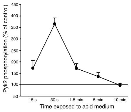
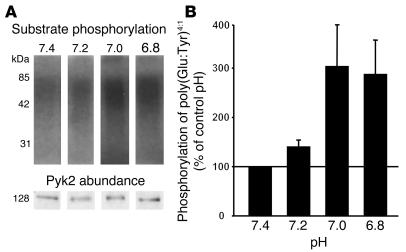
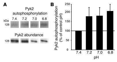
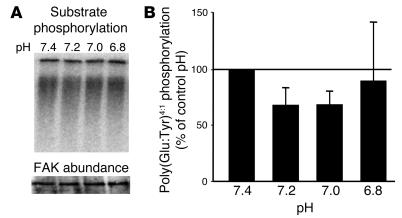
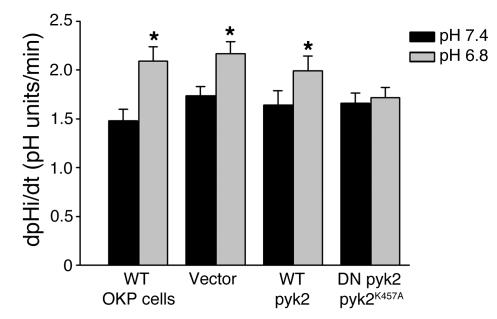
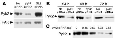
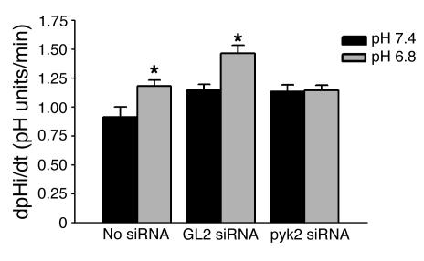
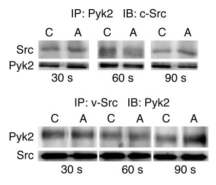
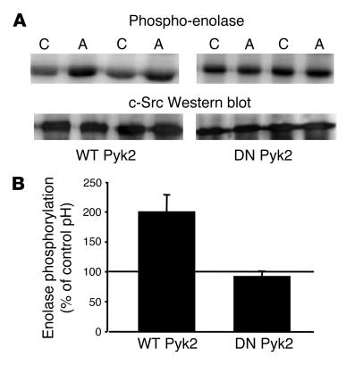
The present study examines the role of Pyk2 in acid regulation of sodium/hydrogen exchanger 3 (NHE3) activity in OKP cells, a kidney proximal tubule epithelial cell line. Incubation of OKP cells in acid media caused a transient increase in Pyk2 phosphorylation that peaked at 30 seconds and increased Pyk2/c-Src binding at 90 seconds. Pyk2 isolated by immunoprecipitation and studied in a cell-free system was activated and phosphorylated at acidic pH. Acid activation of Pyk2 (a) was specific for Pyk2 in that acid did not activate focal adhesion kinase, (b) required calcium, and (c) was associated with increased affinity for ATP. Transfection of OKP cells with dominant-negative pyk2(K457A) or small interfering pyk2 duplex RNA blocked acid activation of NHE3, while neither had an effect on glucocorticoid activation of NHE3. In addition, pyk2(K457A) blocked acid activation of c-Src kinase, which is also required for acid regulation of NHE3. The present results demonstrate that Pyk2 is directly activated by acidic pH and that Pyk2 activation is required for acid activation of c-Src kinase and NHE3. Given that partially purified Pyk2 can be activated by acid in a cell-free system, Pyk2 may serve as the pH sensor that initiates the acid-regulated signaling cascade involved in NHE3 regulation.


Figures

Figure 1
Figure 1
Acid incubation induces Pyk2 phosphorylation in OKP cells. After growing to confluence, cells were rendered quiescent for 48 hours and then exposed to a medium of pH 7.4 (control) or 6.8 (acid) for the indicated time. Pyk2 was then immunoprecipitated and phosphorylation assayed, as described in Methods. Data are plotted as percentage of control; n = 6 for control and acid at each time point. Pyk2 phosphorylation was increased in 4 of 6 experiments (15 seconds), 6 of 6 experiments (30 seconds and 5 minutes), 5 of 6 experiments (90 seconds), and 2 of 6 experiments (10 minutes).
Figure 2
Figure 2
Acid activates Pyk2 in a cell-free system. Wild-type OKP cells were grown to confluence and rendered quiescent for 48 hours. Pyk2 was immunoprecipitated as described in Methods and exposed to buffers at the indicated pH for 5 minutes; kinase activity was assayed as the ability of Pyk2 to phosphorylate poly(Glu-Tyr)4:1. (A) Typical autoradiograph (top) and Western blot (bottom). (B) Summary of the data, which are normalized for Pyk2 abundance and plotted as a percentage of control (pH 7.4); n = 9 for each pH. Pyk2 kinase activity was increased in 8 of 9 experiments at pH 7.2, 7.0, and 6.8.
Figure 3
Figure 3
Acid induces Pyk2 autophosphorylation in a cell-free system. Wild-type OKP cells were grown to confluence and rendered quiescent for 48 hours. Pyk2 was immunoprecipitated as described in Methods and exposed to buffers at the indicated pH for 5 minutes; Pyk2 autophosphorylation was assayed. (A) Typical autoradiograph (top) and Western blot (bottom). (B) Summary of the data, which are normalized for Pyk2 abundance and plotted as a percentage of control (pH 7.4); n = 9 for each pH. Pyk2 phosphorylation was increased in 5 of 9 experiments at pH 7.2 and 7.0 and in 8 of 9 experiments at pH 6.8.
Figure 4
Figure 4
Acid does not activate FAK in a cell-free system. OKP cells were grown to confluence and rendered quiescent for 48 hours. FAK was immunoprecipitated as described in Methods and exposed to buffers at the indicated pH for 5 minutes; kinase activity was assayed as the ability of FAK to phosphorylate poly(Glu-Tyr)4:1. (A) Typical autoradiograph (top) and Western blot (bottom). (B) Summary of the data, which are normalized for FAK abundance and plotted as a percentage of control (pH 7.4); n = 3 for pH 7.4, 7.2, and 7.0, and n = 2 for pH 6.8.
Figure 5
Figure 5
Dominant-negative Pyk2 prevents NHE3 activation by acid. OKP cells were transiently transfected with 1 μg of the indicated DNA for 5 hours, grown to confluence over the next 19 hours, rendered quiescent for 24 hours, and exposed to control (pH 7.4) or acid (pH 6.8) media for 6 hours; Na/H antiporter activity was then assayed as the rate of Na-dependent increase in cell pH. Data are plotted as dpHi/dt, pH units/min; n = 6 (wild-type cells), n = 19 (vector transfected), n = 19 (wild-type Pyk2 transfected), and n = 17 (dominant-negative [DN] Pyk2K457A transfected). *P < 0.01 vs. pH 7.4.
Figure 6
Figure 6
siRNApyk2 decreases Pyk2 protein abundance in OKP cells. Typical blots. (A) Cells were transfected with 1.33 μg/well siRNApyk2 or siRNAGL2 (GL2 siRNA) or transfection reagent only (No siRNA) for 24 hours (at which time they were confluent), rendered quiescent for 24 hours, and harvested. FAK protein abundance was measured for comparison. (B) Cells were transfected with 1.33 μg/well siRNApyk2 for 24 hours (at which time they were confluent) and rendered quiescent. The length of quiescence varied from 0 to 48 hours, so that cells were harvested 24, 48, or 72 hours after the beginning of transfection. (C) Cells were transfected with the indicated amount of siRNApyk2 for 24 hours (at which time they were confluent), rendered quiescent for 24 hours, and harvested.
Figure 7
Figure 7
siRNApyk2 prevents NHE3 activation by acid. OKP cells were transiently transfected with 1.33 μg siRNApyk2 (pyk2 siRNA) per well for 24 hours (at which time they were confluent), rendered quiescent for 18 hours, and exposed to control (pH 7.4) or acid (pH 6.8) medium for 6 hours; then Na/H antiporter activity was assayed. Data are plotted as dpHi/dt, pH units/min. Controls were cells exposed to transfection reagent without siRNA (No siRNA) or cells transfected with a siRNA against a nonmammalian protein (GL2 siRNA); n = 7 (No siRNA), n = 6 (GL2 siRNA), and n = 10 (pyk2 siRNA). * P < 0.05 vs. pH 7.4.
Figure 8
Figure 8
Acid incubation increases Pyk2 and c-Src coimmunoprecipitation. Representative blots. OKP cells transiently transfected with pyk2 and c-Src were grown to confluence, rendered quiescent, and exposed to control (C) (pH 7.4) or acid (A) (pH 6.8) medium for the indicated time; Pyk2 or v-Src was immunoprecipated (IP) as indicated. The immunoprecipitates were divided and immunoblotting (IB) performed using anti-Pyk2 and anti-c-Src antibodies as indicated; n = 3 for each group.
Figure 9
Figure 9
Pyk2 is required for acid activation of c-Src. OKP cells transiently transfected with wild-type or dominant-negative (DN) pyk2 and wild-type c-Src were grown to confluence, rendered quiescent, and exposed to control (pH 7.4) or acid (pH 6.8) medium for 90 seconds; c-Src was immunoprecipitated as described in Methods. c-Src kinase activity was assayed using acid-denatured enolase as the substrate. (A) Typical autoradiograph (top) and Western blot (bottom). (B) Summary of the data, which are normalized for c-Src abundance and plotted as percentage of control (pH 7.4); n = 6 for studies done with wild-type Pyk2 and n = 4 for studies done with the dominant-negative Pyk2. c-Src kinase activity was increased in 6 of 6 experiments in the presence of the wild-type Pyk2 and in 1 of 4 experiments in the presence of the dominant-negative Pyk2.

Comment in

Similar articles

Cited by

References

    1. Alpern RJ, Sakhaee K. The clinical spectrum of chronic metabolic acidosis: homeostatic mechanisms produce significant morbidity. Am. J. Kid. Dis. 1997;29:291–302. - PubMed
    1. Tsuganezawa H, et al. Role of c-Src and ERK in acid-induced activation of NHE3. Kidney Int. 2002;62:41–50. - PubMed
    1. Yamaji Y, Moe OW, Miller RT, Alpern RJ. Acid activation of immediate early genes in renal epithelial cells. J. Clin. Invest. 1994;94:1297–1303. - PMC - PubMed
    1. Yamaji Y, et al. Overexpression of csk inhibits acid-induced activation of NHE-3. Proc. Natl. Acad. Sci. U. S. A. 1995;92:6274–6278. - PMC - PubMed
    1. Yamaji Y, Tsuganezawa H, Moe OW, Alpern RJ. Intracellular acidosis activates c-Src. Am. J. Physiol. 1997;272:C886–C893. - PubMed

Publication types

MeSH terms