

T cell-dependent production of IFN-gamma by NK cells in response to influenza A virus
- PMID: 15599406
- PMCID: PMC535070
- DOI: 10.1172/JCI22797
T cell-dependent production of IFN-gamma by NK cells in response to influenza A virus
Abstract
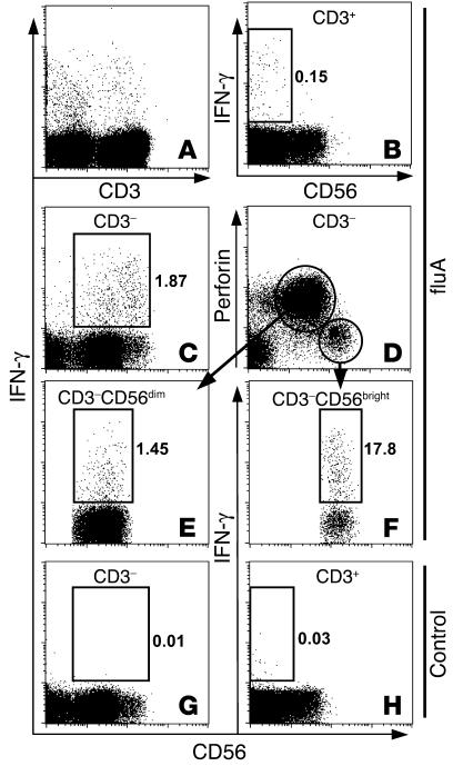
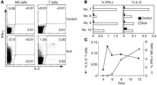
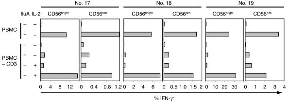
The role of human NK cells in viral infections is poorly understood. We used a cytokine flow-cytometry assay to simultaneously investigate the IFN-gamma response of NK and T lymphocytes to influenza A virus (fluA). When PBMCs from fluA-immune adult donors were incubated with fluA, IFN-gamma was produced by both CD56(dim) and CD56(bright) subsets of NK cells, as well as by fluA-specific T cells. Purified NK cells did not produce IFN-gamma in response to fluA, while depletion of T lymphocytes reduced to background levels the fluA-induced IFN-gamma production by NK cells, which indicates that T cells are required for the IFN-gamma response of NK cells. The fluA-induced IFN-gamma production of NK cells was suppressed by anti-IL-2 Ab, while recombinant IL-2 replaced the helper function of T cells for IFN-gamma production by NK cells. This indicates that IL-2 produced by fluA-specific T cells is involved in the T cell-dependent IFN-gamma response of NK cells to fluA. Taken together, these results suggest that at an early stage of recurrent viral infection, NK-mediated innate immunity to the virus is enhanced by preexisting virus-specific T cells.
Figures


Similar articles
-
Revisiting human natural killer cell subset function revealed cytolytic CD56(dim)CD16+ NK cells as rapid producers of abundant IFN-gamma on activation.Proc Natl Acad Sci U S A. 2011 Jan 11;108(2):728-32. doi: 10.1073/pnas.1012356108. Epub 2010 Dec 27. Proc Natl Acad Sci U S A. 2011. PMID: 21187373 Free PMC article.
-
Invasive Surgery Impairs the Regulatory Function of Human CD56 bright Natural Killer Cells in Response to Staphylococcus aureus. Suppression of Interferon-γ Synthesis.PLoS One. 2015 Jun 19;10(6):e0130155. doi: 10.1371/journal.pone.0130155. eCollection 2015. PLoS One. 2015. PMID: 26090673 Free PMC article.
-
Lipopolysaccharide stimulates the proliferation of human CD56+CD3- NK cells: a regulatory role of monocytes and IL-10.J Immunol. 2000 Jul 1;165(1):139-47. doi: 10.4049/jimmunol.165.1.139. J Immunol. 2000. PMID: 10861046
-
Activation and function of natural killer cell responses during viral infections.Curr Opin Immunol. 1997 Feb;9(1):24-34. doi: 10.1016/s0952-7915(97)80155-0. Curr Opin Immunol. 1997. PMID: 9039782 Review.
-
Dynamic Natural Killer Cell and T Cell Responses to Influenza Infection.Front Cell Infect Microbiol. 2020 Aug 18;10:425. doi: 10.3389/fcimb.2020.00425. eCollection 2020. Front Cell Infect Microbiol. 2020. PMID: 32974217 Free PMC article. Review.
Cited by
-
Sustained effector function of IL-12/15/18-preactivated NK cells against established tumors.J Exp Med. 2012 Dec 17;209(13):2351-65. doi: 10.1084/jem.20120944. Epub 2012 Dec 3. J Exp Med. 2012. PMID: 23209317 Free PMC article.
-
Innate immunity against HIV-1 infection.Nat Immunol. 2015 Jun;16(6):554-62. doi: 10.1038/ni.3157. Nat Immunol. 2015. PMID: 25988887 Review.
-
Elevated gamma interferon-producing NK cells, CD45RO memory-like T cells, and CD4 T cells are associated with protection against malaria infection in pregnancy.Infect Immun. 2008 Apr;76(4):1678-85. doi: 10.1128/IAI.01420-07. Epub 2008 Feb 4. Infect Immun. 2008. PMID: 18250175 Free PMC article.
-
Cutting Edge: Unusual NK cell responses to HIV-1 peptides are associated with protection against maternal-infant transmission of HIV-1.J Immunol. 2009 May 15;182(10):5914-8. doi: 10.4049/jimmunol.0900419. J Immunol. 2009. PMID: 19414742 Free PMC article.
-
Impaired NK Cell Responses to Pertussis and H1N1 Influenza Vaccine Antigens in Human Cytomegalovirus-Infected Individuals.J Immunol. 2015 May 15;194(10):4657-67. doi: 10.4049/jimmunol.1403080. Epub 2015 Apr 8. J Immunol. 2015. PMID: 25855356 Free PMC article.
References
-
- Lamb, R.A., and Krug, R.M. 2001. Orthomyxoviridae: the viruses and their replication. In Fields virology. D.M. Knipe and P.M. Howley, editors. Lippincott Williams & Wilkins. Philadelphia, Pennsylvania, USA. 1533–1579.
-
- Yap KL, Ada GL, McKenzie IF. Transfer of specific cytotoxic T lymphocytes protects mice inoculated with influenza virus. Nature. 1978;273:238–239. - PubMed
-
- McMichael AJ, Gotch FM, Noble GR, Beare PA. Cytotoxic T-cell immunity to influenza. N. Engl. J. Med. 1983;309:13–17. - PubMed
-
- Doherty PC, et al. Effector CD4+ and CD8+ T-cell mechanisms in the control of respiratory virus infections. Immunol. Rev. 1997;159:105–117. - PubMed
Publication types
MeSH terms
Substances
Grants and funding
LinkOut - more resources
Full Text Sources
Other Literature Sources
Research Materials
