Review
. 2005 Apr;54(4):307-14.
doi: 10.1007/s00262-004-0593-x. Epub 2004 Dec 15.

Interaction of PD-L1 on tumor cells with PD-1 on tumor-specific T cells as a mechanism of immune evasion: implications for tumor immunotherapy

Affiliations
Review

Interaction of PD-L1 on tumor cells with PD-1 on tumor-specific T cells as a mechanism of immune evasion: implications for tumor immunotherapy

Christian Blank et al. Cancer Immunol Immunother. 2005 Apr.

Abstract

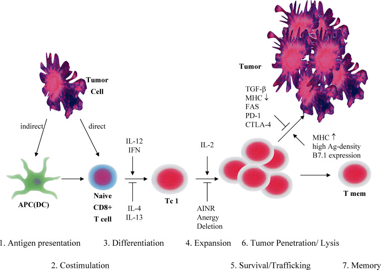
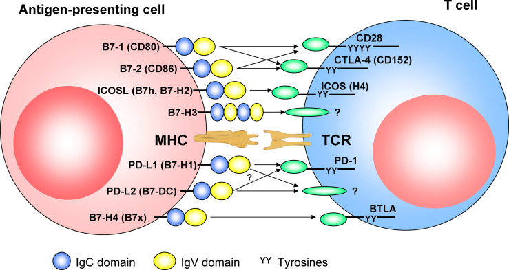
Programmed death receptor ligand 1 (PD-L1, also called B7-H1) is a recently described B7 family member. In contrast to B7-1 and B7-2, PD-L1 does not interact with either CD28 or CTLA-4. To date, one specific receptor has been identified that can be ligated by PD-L1. This receptor, programmed death receptor 1 (PD-1), has been shown to negatively regulate T-cell receptor (TCR) signaling. Upon ligating its receptor, PD-L1 has been reported to decrease TCR-mediated proliferation and cytokine production. PD-1 gene-deficient mice developed autoimmune diseases, which early led to the hypothesis of PD-L1 regulating peripheral tolerance. In contrast to normal tissues, which show minimal surface expression of PD-L1 protein, PD-L1 expression was found to be abundant on many murine and human cancers and could be further up-regulated upon IFN-gamma stimulation. Thus, PD-L1 might play an important role in tumor immune evasion. This review discusses the currently available data concerning negative T-cell regulation via PD-1, the blockade of PD-L1/PD-1 interactions, and the implications for adoptive T-cell therapies.


Figures

Fig. 1
Fig. 1
Schematic description of the requirements for an CD8+ T-cell–mediated antitumor immune response. Naïve tumor-specific CD8+ T cells can be activated directly from tumor cells or indirectly from APCs presenting TAA. In a second step, differentiation toward a type I phenotype is optimal. This step is favored by IL-12 and IFN-γ and suppressed by IL-4 or IL-13. Furthermore, sufficient expansion of tumor-specific T cells is important and may require IL-2. Activation-induced nonresponsiveness (AINR), anergy induction, and T-cell deletion may interfere with sufficient expansion and in vivo survival. Tumor penetration is influenced by chemokines and adhesion molecules. Furthermore, the interaction between tumor-specific T cells and the tumor cell is improved by high MHC expression, high antigen densitiy on the tumor, and B7 expression of the tumor. Immune escape might be mediated via TGF-β, MHC loss, or engagement of Fas, PD-1, or CTLA-4 on the T cell. Finally, long-term responses require the development of a memory immune response.
Fig. 2
Fig. 2
The growing number of B7 superfamily members shaping TCR signaling. CD28 and CTLA-4 have MYPPPY motifs that are essential to bind B7-1 (CD80) and B7-2 (CD86). B7-1 and B7-2 provide important costimulatory signals via an interaction with the constitutively expressed CD28 and provide a negative signal via CTLA-4 (CD152), which is induced upon T-cell activation. ICOS (H4) does not show any binding to B7 and seems to mediate costimulatory effects on recently activated and effector T cells binding to its ligand, ICOS-L (B7 h, B7-H2). B7-H3 is constitutively expressed on IFN-γ–treated DCs and binds to an unidentified receptor on activated, but not on resting, T cells. B7-H3 costimulates proliferation of CD4+ and CD8+ T cells. PD-L1 (B7-H1) negatively regulates proliferation and cytokine production of T and B cells that express PD-1 upon activation. A secondary receptor mediating costimulatory signals from PD-L1 has been postulated, but not yet identified. This receptor possibly interacts preferentially with PD-L2 (B7-DC). B7x (B7-H4) has been reported to interact with BTLA, which is expressed on activated T and B cells, inhibiting lymphocyte activation. In contrast to the APC shown here, most tumor cells lack B-7 costimulatory molecules and express strongly PD-L1, either constitutively or upon IFN-γ. This expression pattern might shift the costimulatory balance of the TCR signal toward inhibition of tumor-specific T cells when interacting with the tumor cell.

Similar articles

Cited by

References

    1. Abbott A. Cancer research: on the offensive. Nature. 2002;416:470–474. - PubMed
    1. Agata Y, Kawasaki A, Nishimura H, Ishida Y, Tsubata T, Yagita H, Honjo T. Expression of the PD-1 antigen on the surface of stimulated mouse T and B lymphocytes. Int Immunol. 1996;8:765–772. - PubMed
    1. Ansari MJ, Salama AD, Chitnis T, Smith RN, Yagita H, Akiba H, Yamazaki T, Azuma M, Iwai H, Khoury SJ, et al. The programmed death-1 (PD-1) pathway regulates autoimmune diabetes in nonobese diabetic (NOD) mice. J Exp Med. 2003;198:63–69. - PMC - PubMed
    1. Bachmann MF, Waterhouse P, Speiser DE, McKall-Faienza K, Mak TW, Ohashi PS. Normal responsiveness of CTLA-4-deficient anti-viral cytotoxic T cells. J Immunol. 1998;160:95–100. - PubMed
    1. Bennett F, Luxenberg D, Ling V, Wang IM, Marquette K, Lowe D, Khan N, Veldman G, Jacobs KA, Valge-Archer VE, et al. Program death-1 engagement upon TCR activation has distinct effects on costimulation and cytokine-driven proliferation: attenuation of ICOS, IL-4, and IL-21, but not CD28, IL-7, and IL-15 responses. J Immunol. 2003;170:711–718. - PubMed