. 2005 Jan;187(1):249-56.
doi: 10.1128/JB.187.1.249-256.2005.

Molecular analysis of the Vibrio cholerae type II secretion ATPase EpsE

Affiliations

Molecular analysis of the Vibrio cholerae type II secretion ATPase EpsE

Jodi L Camberg et al. J Bacteriol. 2005 Jan.

Abstract

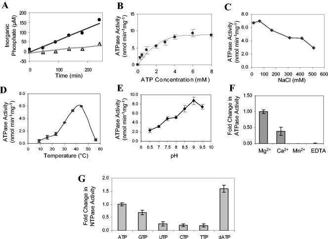
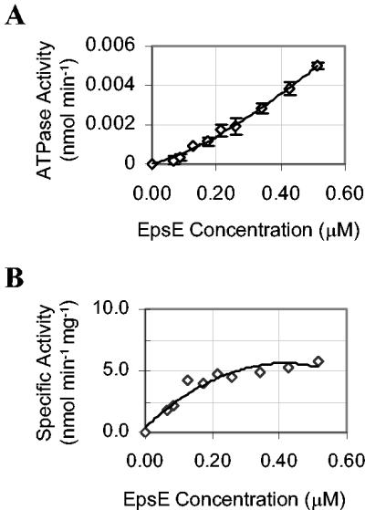
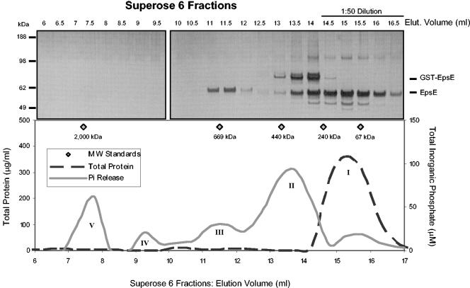
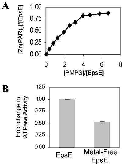
The type II secretion system is a macromolecular assembly that facilitates the extracellular translocation of folded proteins in gram-negative bacteria. EpsE, a member of this secretion system in Vibrio cholerae, contains a nucleotide-binding motif composed of Walker A and B boxes that are thought to participate in binding and hydrolysis of ATP and displays structural homology to other transport ATPases. Here we demonstrate that purified EpsE is an Mg2+-dependent ATPase and define optimal conditions for the hydrolysis reaction. EpsE displays concentration-dependent activity, which may suggest that the active form is oligomeric. Size exclusion chromatography showed that the majority of purified EpsE is monomeric; however, detailed analyses of specific activities obtained following gel filtration revealed the presence of a small population of active oligomers. We further report that EpsE binds zinc through a tetracysteine motif near its carboxyl terminus, yet metal displacement assays suggest that zinc is not required for catalysis. Previous studies describing interactions between EpsE and other components of the type II secretion pathway together with these data further support the hypothesis that EpsE functions to couple energy to the type II apparatus, thus enabling secretion.


Figures

FIG. 1.
FIG. 1.
Purification of EpsE. GST-EpsE was affinity purified from soluble E. coli extract with glutathione Sepharose. EpsE was cleaved from GST by the addition of thrombin (100 U/ml). Purified material was passed over benzamidine Sepharose for removal of thrombin. Fractions were analyzed by reducing SDS-PAGE and stained with GelCode blue. Lanes: 1, insoluble pellet obtained after French press and centrifugation; 2, soluble extract used for purification; 3, column flowthrough; 4 and 5, buffer washes; 6 to 10, fractions obtained by elution with thrombin; 11 and 12, fractions recovered after thrombin removal.
FIG. 2.
FIG. 2.
ATPase activity of EpsE under various conditions. (A) Time course analysis of ATP hydrolysis. EpsE and EpsE(K270A) were purified in parallel and tested for ATPase activity by incubation with 4 mM ATP and 4 mM MgCl2 at 37°C (1 μM final protein concentration). Total Pi was measured at intervals by the malachite green assay. EpsE, closed symbols; EpsE(K270A), open symbols. (B) Concentration of substrate required for enzyme saturation. We tested 0.625 μM EpsE in 100 mM Tris (pH 8.5) for ATPase activity by the addition of increasing amounts of substrate (0 to 8 mM ATP) with an endpoint assay (n = 3 for each substrate concentration). Error is reported as the standard error. (C) ATPase activity for EpsE tested at various NaCl concentrations with an endpoint assay. (D) ATPase activity determination at different temperatures (n = 3). (E) Influence of pH on EpsE ATPase activity (n = 7). (F) Optimal divalent metal to support ATPase activity. The final concentration of divalent metal was 4 mM. Mg2+, n = 14; Ca2+, Mn2+, and EDTA, n = 8. (G) Optimal substrate for hydrolysis. NTPs were added to a final concentration of 4 mM. ATP, GTP, TTP, and dATP, n = 12; CTP, n = 11; UTP, n = 8.
FIG. 3.
FIG. 3.
Concentration-dependent activity of EpsE. (A) ATP hydrolysis was monitored with an endpoint assay at various EpsE concentrations (0.06 to 0.5 μM) with 5 mM MgCl2 and 5 mM ATP at 37°C. Total Pi was measured after 16 h (n = 3 for each enzyme concentration). (B) Concentration-dependent ATP hydrolysis was analyzed on a plot of specific activity (nmol min−1 mg−1) versus EpsE concentration (micromolar).
FIG. 4.
FIG. 4.
Size exclusion chromatography reveals oligomeric forms of EpsE. EpsE was fractionated on a Superose 6 column. Fractions of 0.5 ml were collected and assayed for total protein and ATP hydrolysis. The upper panel shows SDS-PAGE analysis and silver staining of individual fractions. The lower panel shows protein concentration (dashed line) and Pi production (solid line) per fraction elucidating five distinct peaks (I to V). MW, molecular mass; Elut., elution.
FIG. 5.
FIG. 5.
Removal of GST-containing oligomers. EpsE was treated with glutathione Sepharose and applied to a Superose 6 column. Fractions of 0.5 ml were collected and analyzed by SDS-PAGE and silver staining (lower panel). These fractions were compared with the elution profile of untreated EpsE (upper panel). Elut., elution.
FIG. 6.
FIG. 6.
Depletion of divalent metal from EpsE and enzymatic analysis. (A) Increasing concentrations of PMPS were added to a 10 μM solution of EpsE containing 200 μM PAR. Absorbance of PAR was monitored at 500 nm and compared to a ZnCl2 standard curve to measure the amount of divalent metal complexed with PAR upon PMPS addition. (B) ATPase activity of metal-free EpsE was measured with an endpoint assay and compared with that of untreated EpsE. (n = 3).

References

    1. Banecki, B., A. Wawrzynow, J. Puzewicz, C. Georgopoulos, and M. Zylicz. 2001. Structure-function analysis of the zinc-binding region of the Clpx molecular chaperone. J. Biol. Chem. 276:18843-18848. - PubMed
    1. Claret, L., S. R. Calder, M. Higgins, and C. Hughes. 2003. Oligomerization and activation of the FliI ATPase central to bacterial flagellum assembly. Mol. Microbiol. 48:1349-1355. - PMC - PubMed
    1. Collet, J. F., J. C. D'Souza, U. Jakob, and J. C. Bardwell. 2003. Thioredoxin 2, an oxidative stress-induced protein, contains a high affinity zinc binding site. J. Biol. Chem. 278:45325-45332. - PubMed
    1. Donaldson, L. W., U. Wojtyra, and W. A. Houry. 2003. Solution structure of the dimeric zinc binding domain of the chaperone ClpX. J. Biol. Chem. 278:48991-48996. - PubMed
    1. Fekkes, P., J. G. de Wit, A. Boorsma, R. H. Friesen, and A. J. Driessen. 1999. Zinc stabilizes the SecB binding site of SecA. Biochemistry 38:5111-5116. - PubMed

Publication types

MeSH terms

LinkOut - more resources