

Role of the two catalytic domains of DSR-E dextransucrase and their involvement in the formation of highly alpha-1,2 branched dextran
- PMID: 15601714
- PMCID: PMC538823
- DOI: 10.1128/JB.187.1.296-303.2005
Role of the two catalytic domains of DSR-E dextransucrase and their involvement in the formation of highly alpha-1,2 branched dextran
Abstract
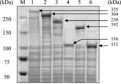
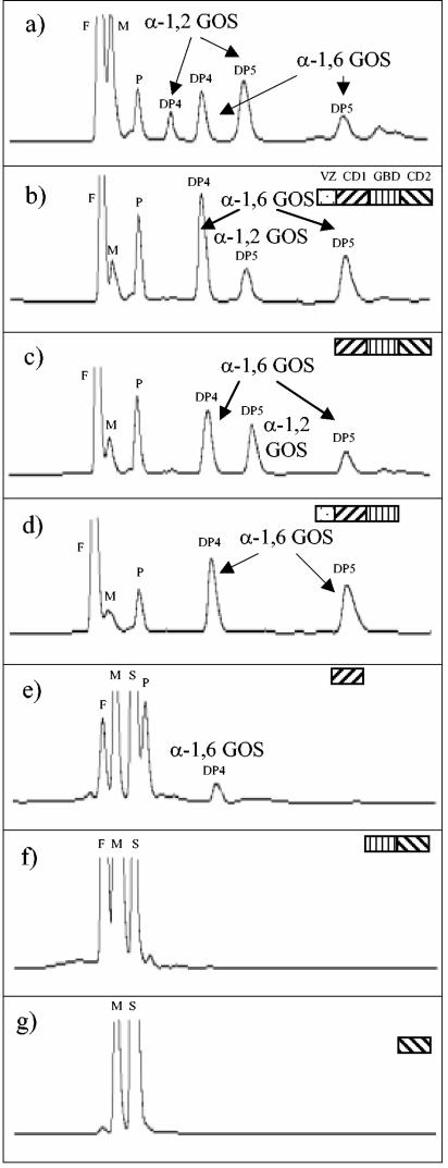
The dsrE gene from Leuconostoc mesenteroides NRRL B-1299 was shown to encode a very large protein with two potentially active catalytic domains (CD1 and CD2) separated by a glucan binding domain (GBD). From sequence analysis, DSR-E was classified in glucoside hydrolase family 70, where it is the only enzyme to have two catalytic domains. The recombinant protein DSR-E synthesizes both alpha-1,6 and alpha-1,2 glucosidic linkages in transglucosylation reactions using sucrose as the donor and maltose as the acceptor. To investigate the specific roles of CD1 and CD2 in the catalytic mechanism, truncated forms of dsrE were cloned and expressed in Escherichia coli. Gene products were then small-scale purified to isolate the various corresponding enzymes. Dextran and oligosaccharide syntheses were performed. Structural characterization by (13)C nuclear magnetic resonance and/or high-performance liquid chromatography showed that enzymes devoid of CD2 synthesized products containing only alpha-1,6 linkages. On the other hand, enzymes devoid of CD1 modified alpha-1,6 linear oligosaccharides and dextran acceptors through the formation of alpha-1,2 linkages. Therefore, each domain is highly regiospecific, CD1 being specific for the synthesis of alpha-1,6 glucosidic bonds and CD2 only catalyzing the formation of alpha-1,2 linkages. This finding permitted us to elucidate the mechanism of alpha-1,2 branching formation and to engineer a novel transglucosidase specific for the formation of alpha-1,2 linkages. This enzyme will be very useful to control the rate of alpha-1,2 linkage synthesis in dextran or oligosaccharide production.
Figures


References
-
- Djouzy, Z., C. Andrieux, V. Pelenc, S. Somarriba, F. Popot, F. Paul, P. Monsan, and O. Szylit. 1995. Degradation and fermentation of α-gluco-oligosaccharides by bacterial strains from human colon: in vitro and in vivo studies in gnotobiotic rats. J. Appl. Bacteriol. 79:117-127. - PubMed
-
- Dols, M., M. Remaud-Simeon, R. M. Willemot, M. R. Vignon, and P. F. Monsan. 1996. Characterization of dextransucrases from Leuconostoc mesenteroides NRRL B-1299. Appl. Biochem. Biotechnol. 62:47-59.
-
- Dols, M., M. Remaud-Simeon, R. M. Willemot, M. R. Vignon, and P. F. Monsan. 1998. Structural characterisation of the maltose acceptor-products synthesised by Leuconostoc mesenteroides NRRL B-1299 dextransucrase. Carbohydr. Res. 305:549-559. - PubMed
MeSH terms
Substances
LinkOut - more resources
Full Text Sources
Other Literature Sources
Molecular Biology Databases
