. 2004 Dec 28;101(52):18024-9.
doi: 10.1073/pnas.0405747102. Epub 2004 Dec 15.

Responses of fibroblasts to anchorage of dorsal extracellular matrix receptors

Affiliations

Responses of fibroblasts to anchorage of dorsal extracellular matrix receptors

Karen A Beningo et al. Proc Natl Acad Sci U S A. .

Abstract

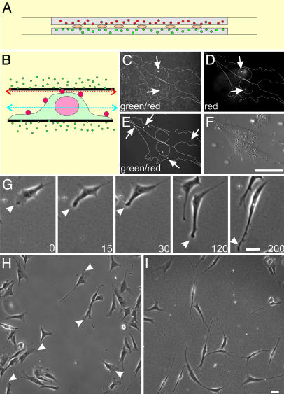
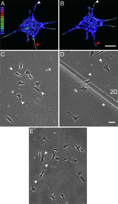
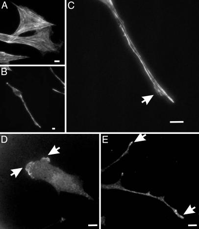
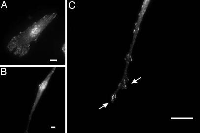
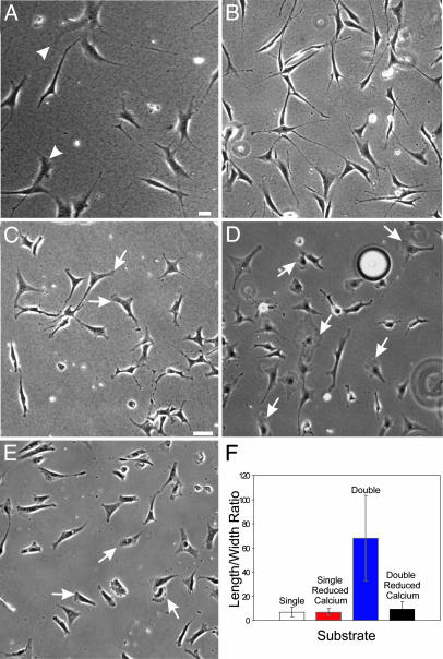
Fibroblasts in 2D cultures differ dramatically in behavior from those in the 3D environment of a multicellular organism. However, the basis of this disparity is unknown. A key difference is the spatial arrangement of anchored extracellular matrix (ECM) receptors to the ventral surface in 2D cultures and throughout the entire surface in 3D cultures. Therefore, we asked whether changing the topography of ECM receptor anchorage alone could invoke a morphological response. By using polyacrylamide-based substrates to present anchored fibronectin or collagen on dorsal cell surfaces, we found that well spread fibroblasts in 2D cultures quickly changed into a bipolar or stellate morphology similar to fibroblasts in vivo. Cells in this environment lacked lamellipodia and large actin bundles and formed small focal adhesions only near focused sites of protrusion. These responses depend on substrate rigidity, calcium ion, and, likely, the calcium-dependent protease calpain. We suggest that fibroblasts respond to both spatial distribution and mechanical input of anchored ECM receptors. Changes in cell shape may in turn affect diverse cellular activities, including gene expression, growth, and differentiation, as shown in numerous previous studies.


Figures

Fig. 1.
Fig. 1.
The double-substrate culturing system and its effects on fibroblast morphology. (A) NIH 3T3 cells are cultured between two sheets of protein-coated polyacrylamide, each ≈75 μm in thickness and embedded with 0.1-μm red (top substrate) or green (bottom substrate) fluorescent microbeads. (B) Cells were surface-labeled with polychromatic beads of 0.5 μm, fixed, and placed in the double substrate with a gap distance of 3.6 μm, to document the initial cell-substrate contact. (C) Confocal image recorded in the green channel and focused slightly underneath the top substrate (red arrow in B) shows both polychromatic beads on the dorsal surface above the nucleus and green beads embedded within the top substrate. (D) Image taken in the red channel on the same focal plane identifies the 0.5-μm polychromatic beads. Beads that bind to more peripheral regions of the dorsal surface are out of focus in this image but are visible on a lower plane of focus (E, taken in the green channel; blue arrow in B), where green beads within the substrates are out of focus. Over a period of 3 h (time in minutes as indicated), long extensions form out of existing lamellipodia (G, arrowhead), such that, at steady state, fibroblasts in the double-substrate culture become highly elongated and lack lamellipodia (I). In contrast, fibroblasts on fibronectin-coated single substrate maintain typical lamellipodia (H, arrowheads). (Bar, 30 μm.)
Fig. 2.
Fig. 2.
Involvement of anchorage of dorsal ECM receptors in morphological changes observed at steady state. NIH 3T3 cells were cultured in the double-substrate system, coated with fibronectin, and embedded with red and green beads in bottom and top substrates, respectively. After overnight incubation, the magnitude of traction stress exerted by the cell on the bottom (A) and top (B) substrate was measured based on displacements of the embedded beads and rendered as color images. Warm (red) colors designate strong traction forces, and cool (blue) colors indicate weaker forces. Red arrows indicate an extension where the cell exerts stronger traction forces on the top substrate than on the bottom substrate. Arrowheads indicate the converse situation for another extension. Color-coded bar represents the magnitude of stress from 1 × 10² to 2.7 × 10⁵ dyne/cm2. (C) When only the bottom sheet is coated with fibronectin, after overnight incubation in the double-substrate culture, cells maintain a morphology similar to those in 2D cultures. (D) In addition, at the boundary between fibronectin-coated single and double substrates, lamellipodia are found where cells make contact with only the bottom substrate, whereas elongated morphology is seen on the double-substrate side (indicated with an asterisk). (E) Cells also maintain a morphology similar to those in 2D cultures when the bottom sheet has been coated with fibronectin and the top sheet has been coated with BSA. (Bar, 30 μm.)
Fig. 3.
Fig. 3.
Organization of actin filaments and Arp3 in fibroblasts cultured in fibronectin-coated 2D and double-substrate cultures. (A) Rhodamine phalloidin staining of actin filaments show many large actin bundles in fibroblasts cultured on 2D polyacrylamide sheets. In contrast, in the double-substrate culture, most actin bundles are located along lateral borders of the extensions (B and C), although there are a few short actin bundles near the nucleus. Arrow indicates a small protrusive region where actin organization is more diffuse. Arp3 staining shows a band of concentration along the leading edge on a fibronectin-coated 2D substrate (D) and discrete foci at the tips of extensions in a double-substrate culture (E). (Bar, 10 μm.)
Fig. 4.
Fig. 4.
Organization of focal adhesions in fibroblasts cultured in fibronectin-coated 2D and double-substrate cultures. Immunofluorescence of paxillin at a low magnification shows many prominent focal adhesions in cells on 2D substrates (A) but few focal adhesions in double-substrate cultures (B). At a higher magnification, small dot-like focal adhesions are observed in an extension, many of which are located near protrusive edges (C, arrows). (Bar, 10 μm.)
Fig. 5.
Fig. 5.
Mechanical input and calcium are required for the response to double-substrate culture. Fibroblasts cultured in a hybrid double substrate of soft (5% acrylamide, 0.06 bis-acrylamide; Young's modulus 1.4 × 10⁴ N/m2) and stiff (5% acrylamide, 0.1% bis-acrylamide; Young's modulus 2.8 × 10⁴ N/m2) polyacrylamide show both elongated morphology and lamellipodia (A, arrowheads). (B) Vehicle-treated control cells in fibronectin-coated double-substrate cultures elongate normally after 2 h in culture. (C and D) In contrast, cells cultured in the presence of 30 μM 5,5′-dimethyl 1,2-bis(2-aminophenoxy)ethane-N,N,N′,N′-tetraacetate acetoxymethyl ester, 3 μM thapsigargin, and 2 mM EGTA maintained a more spread morphology and lamellipodia activities, as indicated by arrows, after 2 h in the double-substrate cultures. (E) Fibroblasts cultured on fibronectin-coated 2D substrates and depleted of calcium maintain lamellipodia (arrows) and show a normal phase morphology. (F) The difference in cell shape is further quantified by measuring the aspect ratio in single- and double-substrate cultures with and without calcium depletion (n = 25 cells, from three to five experiments for each condition). (Bar, 30 μm.)

References

    1. Folkman, J. & Moscona, A. (1978) Nature 273, 345–349. - PubMed
    1. Watt, F. M., Jordan, P. W. & O'Neill, C. H. (1988) Proc. Natl. Acad. Sci. USA 85, 5576–5588. - PMC - PubMed
    1. Dunn, G. A. & Brown, A. F. (1986) J. Cell Sci. 83, 313–340. - PubMed
    1. Curtis, A. S. G. & Wilkinson, C. (1997) Biomaterials 18, 1573–1583. - PubMed
    1. Lo, C.-M., Wang, H.-B., Dembo, M. & Wang, Y.-L. (2000) Biophys. J. 79, 144–152. - PMC - PubMed

Publication types

MeSH terms

LinkOut - more resources