. 2004 Dec 28;101(52):18153-8.
doi: 10.1073/pnas.0405172102. Epub 2004 Dec 15.

Influenza virus entry and infection require host cell N-linked glycoprotein

Affiliations

Influenza virus entry and infection require host cell N-linked glycoprotein

Victor C Chu et al. Proc Natl Acad Sci U S A. .

Abstract

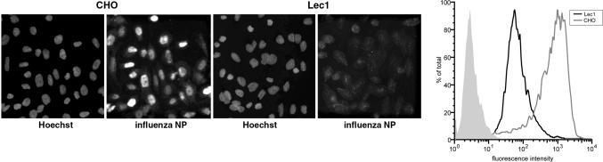
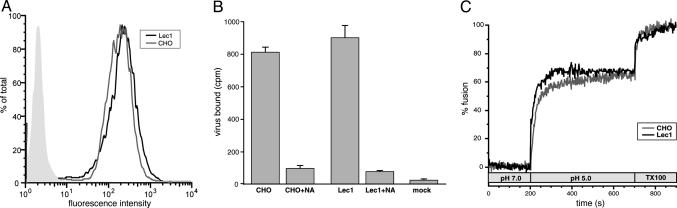
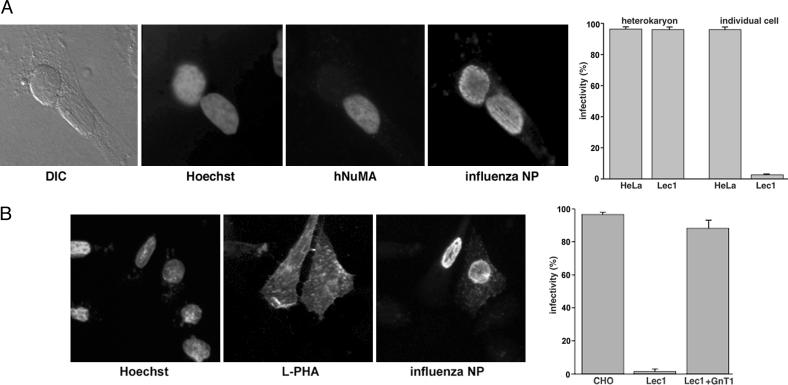
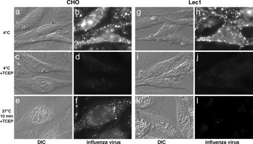
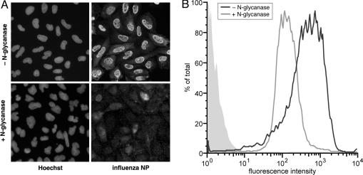
A widely held view of influenza virus infection is that the viral receptor consists of cell surface carbohydrate sialic acid, which can be present as glycoprotein or glycolipid. Here, we examined influenza virus entry and infection in Lec1 cells, a mutant CHO cell line deficient in terminal N-linked glycosylation caused by a mutation in the N-acetylglucosaminyltransferase I (GnT1) gene. We show that influenza virus cannot infect Lec1 cells, despite having full capacity to undergo virus binding and fusion. Lec1 cells also show no virus replication defect, and infection was restored in Lec1 cells expressing wild-type GnT1. Viruses were apparently arrested at the level of internalization from the plasma membrane and were not endocytosed. Lec1 cells were refractory to infection by several strains of influenza virus, including H1 and H3 strains of influenza A, as well as influenza B virus. Finally, cleavage of N-glycans from wild-type CHO cells markedly reduced infection by influenza virus. We suggest that influenza virus specifically requires N-linked glycoprotein for entry into cells, and that sialic acid, although acting as an efficient attachment factor, is not sufficient as an influenza virus receptor in vivo.


Figures

Fig. 1.
Fig. 1.
Lec1 cells are not infected by influenza virus. (Left) CHO and Lec1 cells were infected with influenza virus (WSN) for 5 h and analyzed by immunofluorescence microscopy using anti-NP antibodies, with nuclei counterstained with Hoechst dye 33258. (Right) CHO (gray line) and Lec1 (black line) cells were also analyzed by flow cytometry. Mock-infected Lec1 cells are shown in the filled light gray trace.
Fig. 2.
Fig. 2.
Influenza virus shows no defect in binding or fusion with Lec1 cells. (A) Influenza virus (WSN) was bound to CHO (gray line) and Lec1 (black line) cells at 4°C for 90 min and analyzed by flow cytometry using anti-NP antibodies. Mock treated Lec1 cells are shown in the filled light gray trace. (B) Radioactive influenza virus (WSN) was bound to CHO and Lec1 cells at 4°C for 90 min, washed extensively, and analyzed by scintillation counting. Cells were also pretreated with 5 units/ml neuraminidase (NA) as a control. (C) R18-labeled influenza virus (WSN) was bound to CHO (gray line) and Lec1 (black line) cells at 4°C for 90 min. The temperature was raised to 37°C, and fusion was induced by dropping the pH to 5.0. Fusion was calibrated by induction of complete dequenching with Triton X-100.
Fig. 3.
Fig. 3.
Influenza virus infection of Lec1 cells is rescued in heterokaryons or by expression of GnT1. (A) Heterokaryons were formed by PEG-mediated fusion between Lec1 and HeLa cells and infected with influenza virus (WSN) for 5 h before immunofluorescence microscopy using anti-NP antibodies. Nuclei were counterstained with Hoechst dye 33258, and HeLa cell nuclei were identified by using a human-specific NuMA antibody. Differential interference contrast (DIC) images of the heterokaryons are also shown. For quantitation, we scored the number of Lec1 and HeLa nuclei in heterokaryons that were infected, in comparison to those that we present as individual cells. The numbers of infected Lec1 and HeLa cell nuclei present in heterokaryons or present as individual cells are shown. (B) Lec1 cells were transfected with plasmids encoding the GnT1 gene. Expression was confirmed by the ability of cells to bind FITC-labeled L-PHA lectin. Infection was monitored by immunofluorescence microscopy using anti-NP antibodies and nuclei counterstained with Hoechst dye 33258.
Fig. 4.
Fig. 4.
Lec1 cells are blocked for infection during influenza virus internalization. Biotinylated influenza virus was bound to the surface of CHO and Lec1 cells at 4°C (b and h) for 90 min, and surface biotin was cleaved with TCEP (d and j). Cells were then allowed to internalize virus for 10 min before TCEP treatment (f and l). Cells were analyzed by differential interference contrast (DIC) microscopy or by immunofluorescence microscopy using Alexa Fluor 488–streptavidin.
Fig. 5.
Fig. 5.
Treatment of cells with N-glycanase abrogates influenza virus infection. (A) CHO cells were treated with N-glycanase or untreated and were infected with influenza virus (WSN) for 5 h, followed by analysis by immunofluorescence microscopy using anti-NP antibodies, with nuclei counterstained with Hoechst dye 33258. (B) N-glycanase-treated (black line) and untreated (gray line) cells were also analyzed by flow cytometry. Mock-infected Lec1 cells are shown in the filled light gray trace.

References

    1. Cox, N. J. & Subbarao, K. (2000) Annu. Rev. Med. 51, 407–421. - PubMed
    1. Skehel, J. J. & Wiley, D. C. (2000) Annu. Rev. Biochem. 69, 531–569. - PubMed
    1. Gottschalk, A. (1959) in The Viruses: Biochemical Biological and Biophysical Properties, eds. Burnet, F. M. & Stanley, W. M. (Academic, New York), Vol. 3, pp. 51–61.
    1. Steinhauer, D. A. & Wharton, S. A. (1998) in Textbook of Influenza, eds. Nicholson, K. G., Webster, R. G. & Hay, A. J. (Blackwell Science, Oxford), pp. 54–64.
    1. Suzuki, Y., Ito, T., Suzuki, T., Holland, R. E., Jr., Chambers, T. M., Kiso, M., Ishida, H. & Kawaoka, Y. (2000) J. Virol. 74, 11825–11831. - PMC - PubMed

Publication types

MeSH terms

Substances