Comparative Study
. 2004 Dec 15;18(24):3078-93.
doi: 10.1101/gad.1255704.

Release of initiation factors from 48S complexes during ribosomal subunit joining and the link between establishment of codon-anticodon base-pairing and hydrolysis of eIF2-bound GTP

Affiliations
Comparative Study

Release of initiation factors from 48S complexes during ribosomal subunit joining and the link between establishment of codon-anticodon base-pairing and hydrolysis of eIF2-bound GTP

Anett Unbehaun et al. Genes Dev. .

Abstract

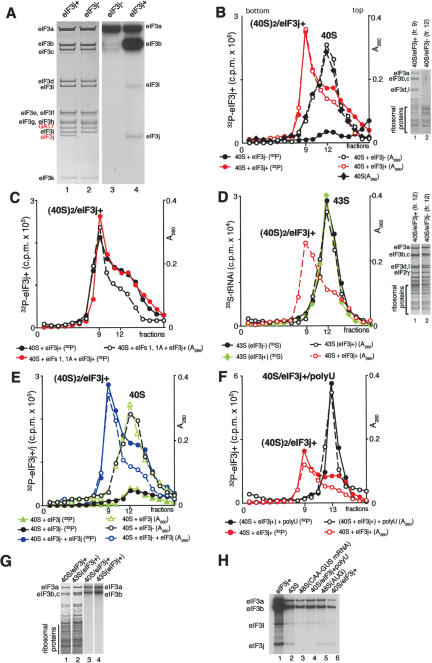
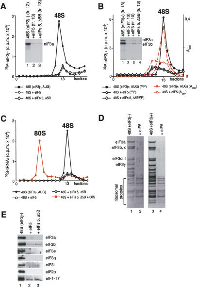
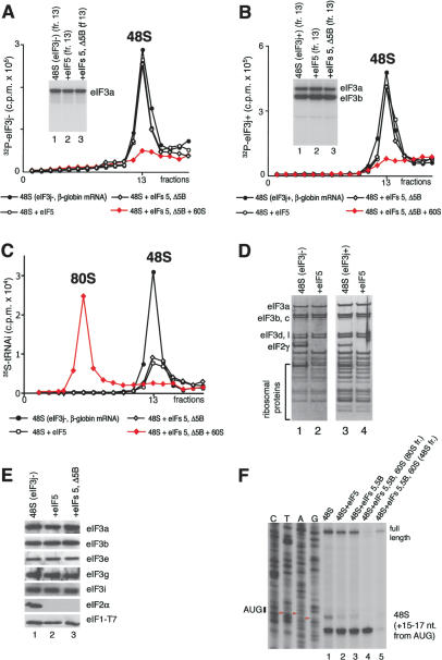
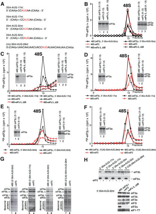
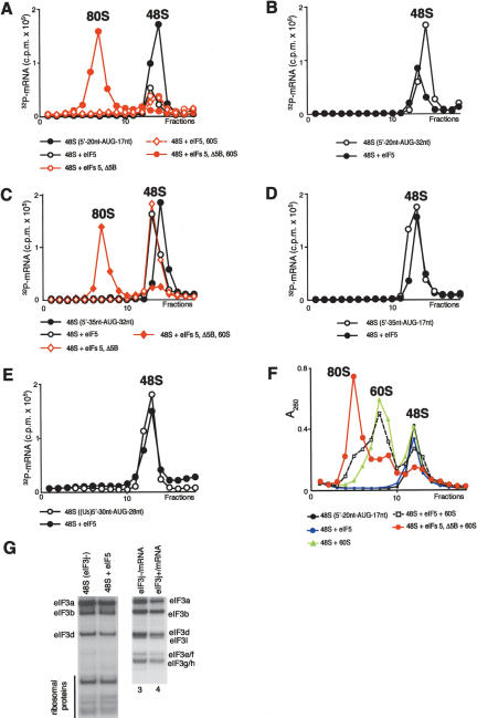
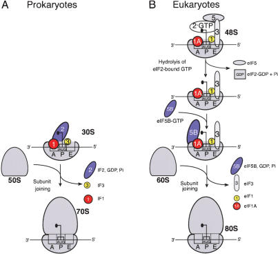
The 40S subunit in 48S complexes formed at the initiation codon of mRNA is bound to eukaryotic initiation factor (eIF) 3, eIF1, eIF1A, and an eIF2/GTP/Met-tRNAi(Met) ternary complex and can therefore not join a 60S subunit directly to form an 80S ribosome. We report that eIF5-induced hydrolysis of eIF2-bound GTP in 48S complexes led to release of eIF2-GDP but not eIF3 or eIF1. eIF5B did not influence factor release in the absence of 60S subunits. Therefore eIF3 and eIF1 dissociate from 40S subunits during, rather than before, the eIF5B-mediated subunit joining event. In the absence of eIF1, eIF5-stimulated hydrolysis of eIF2-bound GTP occurred at the same rate in 43S pre-initiation and 48S initiation complexes. GTP hydrolysis in 43S complexes assembled with eIF1 was much slower than in 43S or 48S complexes assembled without eIF1. Establishment of codon-anticodon base-pairing in 48S complexes relieved eIF1's inhibition. Thus, in addition to its role in initiation codon selection during 48S complex formation, eIF1 also participates in maintaining the fidelity of the initiation process at a later stage, hydrolysis of eIF2-bound GTP, by inhibiting premature GTP hydrolysis and by linking establishment of codon-anticodon base-pairing with GTP hydrolysis.


Figures

Figure 2.
Figure 2.
Subunit composition of eIF3, phosphorylation of eIF3 by cAMP-dependent kinase, and eIF3's binding to 40S subunits. (A) Coomassie staining of HeLa eIF3j+ and eIF3j- and autoradiography of eIF3j- and eIF3j+ 32P-phosphorylated by cAMP-dependent kinase. eIF3 subunits are indicated. (B,C) Binding of 32P-eIF3j+ and 32P-eIF3j- to 40S subunits in the absence (B) and presence (C) of eIF1 and eIF1A, assayed by sucrose density gradient centrifugation and analyzed by optical density, Cerenkov counting (B,C) and gel electrophoresis (B, right). (D) 43S complex formation in the presence of 40S subunits, eIF2, [35S]Met-formula image, eIF1, eIF1A, and eIF3j+ or eIF3j- assayed by sucrose density gradient centrifugation and analyzed by optical density, scintillation counting, and gel electrophoresis (right). The optical density of eIF3j+/40S subunit complexes subjected to sucrose density gradient centrifugation is included for comparison of their mobility and that of 43S complexes. (E) Influence of rec-eIF3j subunit on binding of 32P-eIF3j- to 40S subunits and binding of 32P-eIF3j itself to 40S subunits assayed by sucrose density gradient centrifugation and analyzed by optical density and Cerenkov counting. (F) Influence of poly(U) RNA on binding of 32P-eIF3j+ to 40S subunits assayed by sucrose density gradient centrifugation and analyzed by optical density and Cerenkov counting. Sedimentation was from right to left in all cases. Ribosomal complexes are indicated above appropriate peaks. Upper fractions from sucrose gradients have been omitted for clarity. (G) Comparison of the relative amounts of 40S subunits and 32P-eIF3j+ in binary eIF3j+/40S subunit complexes and in 43S complexes separated by sucrose density gradient centrifugation and analyzed by fluorescent SYPRO staining (lanes 1,2) and autoradiography (lanes 3,4) after gel electrophoresis. (H) Presence of the eIF3j subunit from 32P-eIF3j+ in different ribosomal complexes isolated by sucrose density gradient centrifugation.
Figure 1.
Figure 1.
Influence of initiation factors and codon-anticodon base-pairing on hydrolysis of eIF2-bound GTP. 43S complexes were assembled from 40S subunits, eIF1A, eIF3, and eIF2/[32P]GTP/formula image ternary complex; purified by sucrose density gradient centrifugation; and incubated with AUG, UUG, CAA triplets, mRNAs, and eIF1, eIF1A, eIF4A, eIF4B, eIF4F, eIF5, and ΔeIF5B as indicated. GTP hydrolysis was assayed by release of 32Pi. Data represent the average of five or more experiments.
Figure 3.
Figure 3.
Factor release from 48S complexes assembled on AUG triplets after hydrolysis of eIF2-bound GTP. (A) Release of 32P-eIF3j- and (B) 32P-eIF3j+ from 48S complexes assembled on AUG triplets in the presence of eIF2, eIF3, eIF1, and eIF1A, incubated with eIF5 and ΔeIF5B, as indicated, and assayed after sucrose density gradient centrifugation by Cerenkov counting, optical density measurement, and gel electrophoresis/autoradiography of peak fractions (inset panels). (C) Association of [35S]Met-formula image with ribosomal complexes formed after incubating 48S complexes with eIF5, ΔeIF5B, and 60S subunits, as indicated, and separated by sucrose density gradient centrifugation. Upper fractions of gradients have been omitted for clarity. Ribosomal complexes are indicated above appropriate peaks. (D,E) Association of eIF1, eIF2, eIF3j-, and eIF3j+ with 40S subunits before and after treatment with eIF5 and ΔeIF5B of 48S complexes assembled on AUG triplets, analyzed by fluorescent SYPRO staining (D) or immunoblotting (E), after gel electrophoresis of peak fractions obtained after sucrose density gradient centrifugation of ribosomal complexes. eIF2 and eIF3 subunits, eIF1, and ribosomal proteins are indicated.
Figure 4.
Figure 4.
Release of initiation factors from 48S complexes assembled on β-globin mRNA after hydrolysis of eIF2-bound GTP. (A,B) Association of 32P-eIF3j-(A) and 32P-eIF3j+ (B) with 48S complexes assembled on β-globin mRNA in the presence of eIF2, eIF3, eIF1, eIF1A, eIF4A, eIF4B, and eIF4F before and after incubation with eIF5, ΔeIF5B, and 60S subunits, as indicated, and assayed after sucrose density gradient centrifugation by Cerenkov counting and gel electrophoresis/autoradiography of peak fractions (inset panels). (C) Association of [35S]Met-formula image with ribosomal complexes formed after incubating 48S complexes with eIF5, ΔeIF5B, and 60S subunits, as indicated, and separated by sucrose density gradient centrifugation. Upper fractions of gradients have been omitted for clarity. Ribosomal complexes are indicated above appropriate peaks. (D,E) Association of eIF1, eIF2, eIF3j- and eIF3j+ with 40S subunits before and after treatment with eIF5 and ΔeIF5B of 48S complexes assembled on β-globin mRNA, analyzed by fluorescent SYPRO staining (D) or immunoblotting (E), after gel electrophoresis of peak fractions obtained after sucrose density gradient centrifugation of ribosomal complexes. eIF2 and eIF3 subunits, eIF1, and ribosomal proteins are indicated. (F) Association of β-globin mRNA with 48S complexes before and after incubation with eIF5, eIF5, and ΔeIF5B, or eIF5, ΔeIF5B, and 60S subunits, and association with 80S ribosomes formed by incubating 48S complexes with eIF5, ΔeIF5B, and 60S subunits, indicated by toeprints 15-17 nt from the initiation codon. The red arrowheads indicate the positions of A, U, and G nucleotides of the β-globin mRNA initiation codon.
Figure 5.
Figure 5.
Association of eIF3 with 40S subunits before and after hydrolysis of eIF2-bound GTP in 48S complexes assembled on mRNAs with different lengths. (A) mRNA sequences. (B-F) Release of 32P-eIF3j- and 32P-eIF3j+ from 48S complexes assembled with eIF1, eIF1A, eIF2, and eIF3 on 35nt-AUG-32nt (B), 20nt-AUG-17nt (C), 35nt-AUG-17nt (D), 20nt-AUG-32nt (E), and (U5)-30nt-AUG-28nt (F) mRNAs after incubation with eIF5 or eIF5 and ΔeIF5B, as indicated, and separated by sucrose density gradient centrifugation, followed by Cerenkov counting and autoradiography after gel electrophoresis of peak fractions (inset panels). (G,H) Association of eIF1, eIF2, and eIF3j- with 40S subunits before and after treatment with eIF5 and ΔeIF5B of 48S complexes assembled on different mRNAs (as indicated), analyzed by fluorescent SYPRO staining (G) or immunoblotting (H), after gel electrophoresis of peak fractions obtained after sucrose density gradient centrifugation of ribosomal complexes. eIF2 and eIF3 subunits, eIF1, and ribosomal proteins are indicated.
Figure 6.
Figure 6.
Association of mRNA with 40S subunits and 80S ribosomes before and after treatment with eIF5, ΔeIF5B, and 60S subunits of 48S complexes assembled on 20nt-AUG-17nt (A), 20nt-AUG-32nt (B), 35nt-AUG-32nt (C), 35nt-AUG-17nt (D), and (U5)-30nt-AUG-28nt (E) mRNAs, as indicated. Association of 32P-mRNA with ribosomal complexes was assayed by Cerenkov counting after sucrose density gradient centrifugation. (F) 80S complex formation after treatment with eIF5, ΔeIF5B, and 60S subunits (as indicated) of 48S complexes assembled on 20nt-AUG-17nt mRNA. Ribosomal complexes were separated by sucrose density gradient centrifugation and assayed by measuring optical density. Ribosomal complexes are indicated above appropriate peaks. Upper fractions from gradients have been omitted for clarity. (G) UV cross-linking of 32P-(U5)-30nt-AUG-28nt mRNA to components of sucrose density gradient-purified 48S complexes before and after induction of hydrolysis of eIF2-bound GTP and in binary complexes with eIF3j- and eIF3j+. UV cross-linked eIF3 subunits and ribosomal proteins are indicated.
Figure 7.
Figure 7.
Comparative ribosomal subunit joining model for prokaryotes (A) and eukaryotes (B).

References

    1. Asano K.S., Phan, L., Anderson, J., and Hinnebusch, A.G. 1998. Complex formation by all five homologues of mammalian translation initiation factor 3 subunits from Saccharomyces cerevisiae. J. Biol. Chem. 273: 18573-18585. - PubMed
    1. Benne R. and Hershey, J.W.B. 1976. Purification and characterization of initiation factor IF-E3 from rabbit reticulocytes. Proc. Natl. Acad. Sci. 73: 3005-3009. - PMC - PubMed
    1. ____. 1978. The mechanism of action of protein synthesis initiation factors from rabbit reticulocytes. J. Biol. Chem. 253: 3078-3087. - PubMed
    1. Carter A.P., Clemons, W.M., Brodersen, D.E., Morgan-Warren, R.J., Hartsch., T., Wimberly, B.T., and Ramakrishnan, V. 2001. Crystal structure of an initiation factor bound to the 30S ribosomal subunit. Science 291: 498-501. - PubMed
    1. Cayama E., Yepez, A., Rotondo, F., Bandeira, E., Ferreras, A.C., and Triana-Alonso, F.J. 2000. New chromatographic and biochemical strategies for quick preparative isolation of tRNA. Nucleic Acids Res. 28: E64. - PMC - PubMed

Publication types

MeSH terms

LinkOut - more resources