. 2005 Jan;25(1):44-59.
doi: 10.1128/MCB.25.1.44-59.2005.

Molecular mechanism of hTid-1, the human homolog of Drosophila tumor suppressor l(2)Tid, in the regulation of NF-kappaB activity and suppression of tumor growth

Affiliations

Molecular mechanism of hTid-1, the human homolog of Drosophila tumor suppressor l(2)Tid, in the regulation of NF-kappaB activity and suppression of tumor growth

Hua Cheng et al. Mol Cell Biol. 2005 Jan.

Abstract

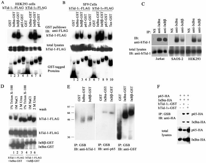
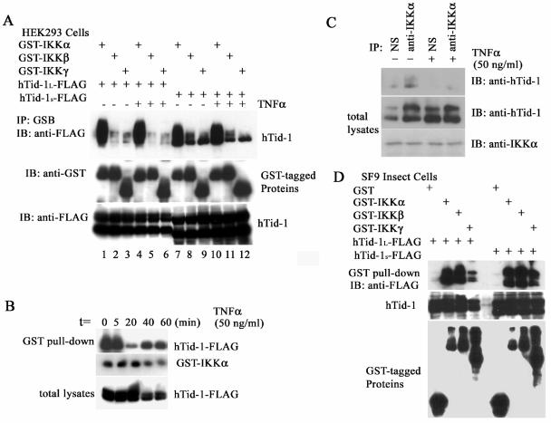
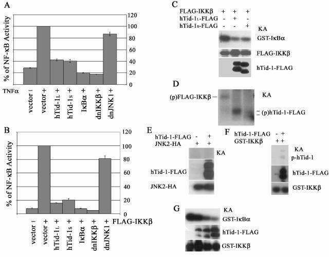
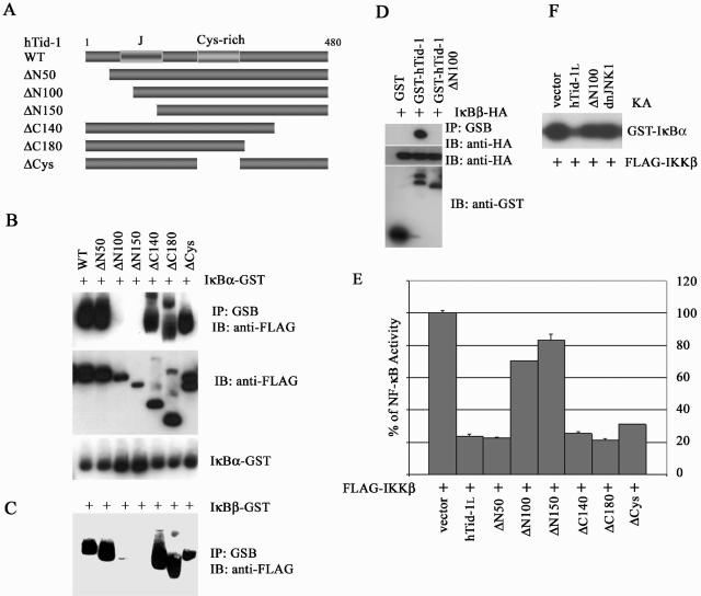
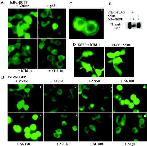
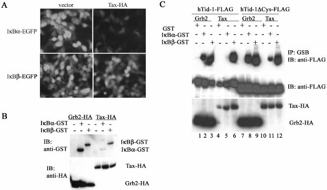
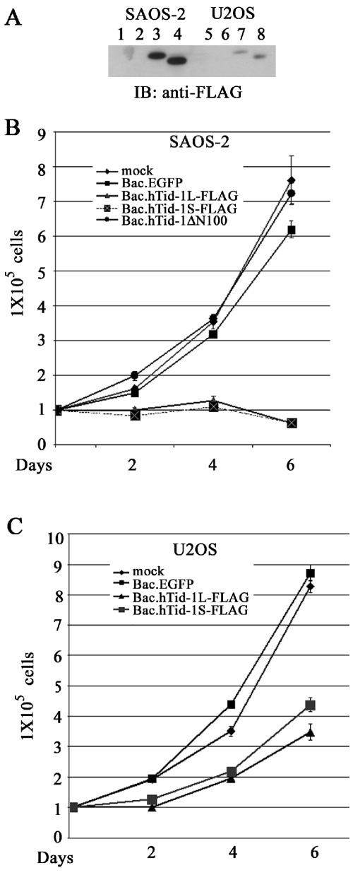
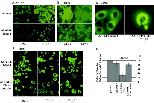
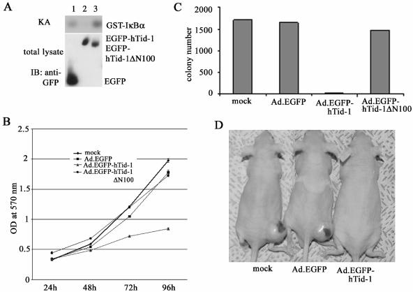
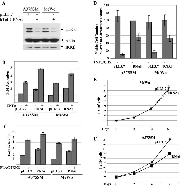
hTid-1, a human homolog of the Drosophila tumor suppressor l(2)Tid and a novel DnaJ protein, regulates the activity of nuclear factor kappaB (NF-kappaB), but its mechanism is not established. We report here that hTid-1 strongly associated with the cytoplasmic protein complex of NF-kappaB-IkappaB through direct interaction with IkappaBalpha/beta and the IKKalpha/beta subunits of the IkappaB kinase complex. These interactions resulted in suppression of the IKK activity in a J-domain-dependent fashion and led to the cytoplasmic retention and enhanced stability of IkappaB. Overexpression of hTid-1 by using recombinant baculovirus or adenovirus led to inhibition of cell proliferation and induction of apoptosis of human osteosarcoma cells regardless of the p53 expression status. Adherent cultured cells transduced with Ad.hTid-1 detached from the dish surface. Morphological changes consistent with apoptosis and cell death were evident 48 h after Ad.EGFP-hTid-1 transduction. In contrast, cells transduced with Ad.EGFP or Ad.EGFP-hTd-1DeltaN100, a mutant that has the N-terminal J domain deletion and that lost suppressive activity on IKK, continued to proliferate. Similar data were obtained with A375 human melanoma cells. Ad.EGFP or Ad.EGFP-hTd-1DeltaN100 ex vivo-transduced A375 cells injected subcutaneously into nude mice produced growing tumors, whereas Ad.EGFP-hTid-1-transduced cells did not. Collectively, the data suggest that hTid-1 represses the activity of NF-kappaB through physical and functional interactions with the IKK complex and IkappaB and, in doing so, it modulates cell growth and death.


Figures

FIG. 1.
FIG. 1.
hTid-1 associates with the cytoplasmic NF-κB-IκB protein complex through direct binding to IκBα and IκBβ. (A) hTid-1 interacts with IκBα and IκBβ in cotransfected HEK293 cells. Transient cotransfections were performed as described in Materials and Methods with hTid-1-FLAG and GST fusion constructs including IκBα-GST, IκBβ-GST, and p65-GST (1 μg each). The GST pull-down precipitates were analyzed by anti-FLAG immunoblot (top panel). The membrane was then stripped and reblotted with anti-GST (bottom panel). Total cellular extracts were analyzed with anti-FLAG immunoblot to detect hTid-1-FLAG protein levels (middle panel). (B) hTid-1 coprecipitates with IκB proteins expressed in SF9 insect cells. In the top panel, a GST pull-down was analyzed by anti-FLAG immunoblotting. The GST fusion proteins in the GST pull-down were detected by using anti-GST antibody (bottom panel), and the total lysates were examined for hTid-1 expression levels with anti-FLAG (middle panel). (C) Protein complex formation of endogenous hTid-1 and IκB proteins. Endogenously expressed hTid-1 appeared in the anti-IκBα and anti-IκBβ immunoprecipitates but not in the nonspecific immunoprecipitates in Jurkat T, SAOS-2, and HEK293 cells. The immunoprecipitates were analyzed with anti-hTid-1 immunoblot (upper panel), and the total protein lysates were assessed for endogenous hTid-1 protein levels (lower panel). (D) hTid-1 binds to IκB proteins with high affinity. SF9 cells (2 × 10⁶) were coinfected with recombinant baculoviruses coexpressing hTid-1S-FLAG and IκBα-GST or IκBβ-GST (100 μl of P3 virus stocks). The GST pull-down precipitates were washed extensively with 1% Triton X-100 lysis buffer (lanes 1 and 4), 1 M NaCl (lanes 2 and 5) or 2 M urea (lanes 3 and 6) in addition to 1% Triton X-100. The precipitates were then analyzed by immunoblotting with anti-FLAG antibody (top panel), and the membrane was stripped and reblotted with anti-GST (bottom panel). The hTid-1S-FLAG expression levels are indicated in the middle panel. (E) Endogenous hTid-1 and p65 proteins were coprecipitated with IκBα-GST and IκBβ-GST proteins. HEK cells (2 × 10⁶ cells) were transfected with GST, IκBα-GST, or IκBβ-GST. The GST pull-down precipitates were evaluated with anti-hTid-1 antibody (left panel). The membrane was stripped and reblotted with anti-p65 antibody (middle panel); the GST fusion protein levels are indicated in the right panel. (F) hTid-1 is associated with the p65-IκBα protein complex. HA-tagged IκBα and p65, with or without hTid-1L-GST, were cotransfected into HEK293 cells. The GST pull-down was analyzed by anti-HA blotting (upper panel), and the IκBα-HA and p65-HA expression levels in total lysates were assessed by anti-HA blotting (lower panel). The results are representative ones of three independent experiments.
FIG. 2.
FIG. 2.
Both hTid-1 variants bind to the IKK complex and repress kinase activity. (A) hTid-1 also associates with IKKα/β. Transient cotransfection of FLAG-tagged hTid-1 with GST-tagged subunits of the IKK complex (1 μg each) was performed by using a method similar to that described in Fig. 1. The transfected cells were stimulated with or without TNF-α (20 ng/ml, 20 min). The GST pull-down precipitates were analyzed with anti-FLAG immunoblot (top panel). The amount of GST fusion proteins is indicated at the middle panel, whereas the hTid-1-FLAG protein levels are shown at the bottom panel. (B) Time course of the interaction of hTid-1 and IKKα upon cell stimulation with TNF-α. Cellular lysates from hTid-1-IKKα (0.2 μg each)-cotransfected HEK cells at different times of TNF-α stimulation (50 ng/ml) were assessed for the coprecipitation of both coexpressed proteins. In the top panel, hTid-1 proteins were detected by a GST pull-down assay with an anti-FLAG antibody. The expression levels of GST-IKKα and hTid-1-FLAG are shown in the middle and bottom panels, respectively. (C) Interaction of hTid-1 and IKKα with endogenously expressed proteins. Total lysates from HEK293 cells stimulated with or without TNF-α (50 ng/ml) were immunoprecipitated by nonspecific antibody (anti-FLAG) or by anti-IKKα. The immunoprecipitates were then analyzed by anti-hTid-1 blotting (top panel). The endogenous proteins of hTid-1 and IKKα from total lysates are shown in the middle and bottom panels, respectively. (D) hTid-1 binds directly to the subunits of IKKα and IKKβ. Coprecipitation of hTid-1-FLAG by GST tagged three subunits of the IKK complex expressed in SF9 insect cells. The top panel shows that the hTid-1-FLAG protein was coprecipitated by GST-tagged IKK subunits, the middle panel shows the hTid-1-FLAG expression level from the total lysates, and the GST-tagged IKK subunits from the GST pull-down is shown in the bottom panel.
FIG. 3.
FIG. 3.
hTid-1 downregulates TNF-α-mediated activation of NF-κB and represses the kinase activity of IKK. Both variants of hTid-1 suppress TNF-α (A)- and IKKβ (B)-induced activation of NF-κB by the NF-κB reporter assay, as described previously (8). The results shown are averages ± the SD; experiments were repeated three times with similar patterns. (C) Both hTid-1 variants inhibit serine phosphorylation of IκBα by IKKβ. The in vitro kinase assay was performed as described in Materials and Methods. The top panel shows the in vitro phosphorylated GST-IκBα; the protein levels of FLAG-IKKβ in the total cellular extracts (middle panel) were detected by anti-IKK immunoblotting, and the hTid-1 protein expression shown in the bottom panel was analyzed with anti-FLAG blot. KA, kinase assay. (D) Kinase-active IKKβ phosphorylates hTid-1. Transient cotransfection of FLAG-IKKβ and hTid-1-FLAG was performed in HEK293 cells. The FLAG-tagged proteins were immunoprecipitated with anti-FLAG antibody plus protein A beads. In vitro kinase assay was performed by using conditions similar to those described above with the exception that GST-IκBα protein was not added to the kinase reaction. The phosphorylated IKKβ and hTid-1 proteins are indicated (p, phosphorylated protein). (E) hTid-1 is not phosphorylated by JNK2. Transient cotransfection of hTid-1L-FLAG and JNK2-HA in HEK293 cells were performed, and the total lysates were coimmunoprecipitated by both anti-FLAG and anti-HA. The in vitro kinase assay was done by the same condition as the IKK kinase reaction. Expressions of hTid-1L-FLAG and JNK2 from total lysates are indicated. (F) Phosphorylation of hTid-1 by IKKβ by using recombinant proteins expressed in SF9 insect cells. (G) hTid-1 suppresses IKKβ activity in SF9 insect cells. The fixed amount of Bac.GST-IKKβ (100 μl) and various amounts of Bac.hTid-1L-FLAG (0, 10, 50, or 200 μl) were used to coinfect SF9 cells (5 × 10⁶ cells). An in vitro kinase assay was performed to detect GST-IκBα phosphorylation (top panel). Protein expression levels of hTid-1-FLAG and GST-IKKβ are shown in the middle and bottom panels, respectively.
FIG. 4.
FIG. 4.
An N-terminal domain of hTid-1 is required for interaction with IκB and is important for hTid-1's repression of NF-κB activity. (A) Schematics of the wild-type and deletion mutants of hTid-1. The N-terminal conserved signature J domain and the central Cys-rich domain are indicated. The in vivo interaction of wild-type hTid-1 and its mutant proteins with GST-tagged IκBα (B) or IκBβ (C) were evaluated by using transient cotransfections in HEK cells as described in Fig. 1A. The presence of FLAG-tagged hTid-1 proteins in the GST pull-down was analyzed by anti-FLAG immunoblotting (B, top panel), and the membrane was stripped and reblotted with anti-GST (B, bottom panel). The expression of various hTid-1 proteins in whole-cell extracts was detected with anti-FLAG immunoblot (B, middle panel). Only hTid-1 proteins coprecipitated with IκBβ-GST are shown in panel C. (D) The in vivo binding of GST-tagged hTid-1 or the ΔN100 mutant with IκBβ-HA. In the top panel, proteins in the GST pull-down were analyzed with anti-HA antibody. The middle panel shows the protein levels of HA-tagged IκBβ; GST fusion proteins were detected by anti-GST immunoblotting (bottom panel). (E) The N-terminal domain of hTid-1 is important for the NF-κB repressive activity of the hTid-1. The repressive activity of the hTid-1 mutants was assessed by using the NF-κB reporter assay. The results shown are averages ± the SD; experiments were repeated three times with similar patterns. (F) In vitro kinase assay was performed by cotransfection of the kinase active IKKβ with hTid-1 or with the ΔN100 mutant in HEK cells, and the kinase activity was examined as previously described (8).
FIG. 5.
FIG. 5.
hTid-1 retains IκBα in the cytoplasm. (A) hTid-1 blocks the nuclear import of free IκBα. EGFP-tagged IκBα (1 μg) was cotransfected with empty vector, p65-HA, hTid-1L-FLAG, or hTid-1S-FLAG (1 μg each) in HEK293 cells. The fluorescent signal was examined with a fluorescence microscope 24 h after transfection. (B) Analysis of the subcellular distribution and expression of IκBα-EGFP-cotransfected wild-type hTid-1 and various hTid-1 mutants in HEK cells as indicated in the figure. (C) Panel B4 was enlarged for better viewing and showed protein aggregation and plasma and nuclear membrane attachment of IκBα-EGFP-cotransfected with the ΔN100 mutant. The results represent at least three independent experiments. (D) hTid-1 has no effect on EGFP subcellular distribution. EGFP was cotransfected with hTid-1-FLAG or the ΔN100 mutant in HEK cells. The EGFP fluorescence intensity and cellular distribution were not altered by coexpression with hTid-1. (E) Anti-GFP immunoblot to detect IκBα-EGFP levels from total lysates in the HEK293 cells that were cotransfected with hTid-1L-FLAG or ΔN100.
FIG. 6.
FIG. 6.
hTid-1 protects IκBα from degradation induced by an IKK activator, Tax. (A) Visualization of EGFP-tagged IκBα degradation induced by Tax. IκBα-EGFP or IκBβ-EGFP (1 μg) was cotransfected with an empty vector or with Tax1-HA (1 μg) in HEK293 cells. The fluorescence images were taken 24 h after transfection. (B) Immunoblot assay of IκBα-induced degradation by Tax. GST-tagged IκBα or IκBβ was cotransfected with Grb2-HA (a negative control) or with Tax-HA in HEK293 cells. The total protein extracts were analyzed with immunoblot with anti-GST (upper panel). Expression of the HA-tagged Grb2 and Tax is shown in the lower panel. (C) A Tax binding-minus mutant of hTid-1 (hTid-1ΔCys) protects the hTid-1-IκBα complex from dissociation induced by Tax. Wild-type hTid-1-FLAG or hTid-1ΔCys-FLAG was cotransfected with GST-tagged IκBα or IκBβ, together with Grb2-HA or Tax-HA, into HEK cells. Protein precipitates from a GST pull-down were analyzed by anti-FLAG immunoblotting (top panel), and the total cellular protein extracts were examined with anti-FLAG blot to determine FLAG-tagged hTid-1 expression (middle panel). Protein levels of HA-tagged Grb2 and Tax are shown in the bottom panel.
FIG. 7.
FIG. 7.
Overexpression of hTid-1 induces cell growth arrest and death. (A) Expression of the hTid-1 variants by using recombinant baculoviruses to transduce SAOS-2 and U2OS cells. The osteosarcoma cells were transduced with baculoviruses expressing EGFP (control), hTid-1L-FLAG, or hTid-1S-FLAG. At 40 h after transduction, the total protein lysates were collected for analysis of the protein expression of hTid-1 with anti-FLAG immunoblot. Lanes 1 to 4, SAOS-2 cells; lanes 5 to 8, U2OS cells. Lanes 1 and 5, mock treatment; lanes 2 and 6, Bac.EGFP; lanes 3 and 7, Bac.hTid-1L-FLAG; lanes 4 and 8, Bac.hTid-1S-FLAG. (B) Growth rate of SAOS-2 cells (105 cells) transduced with Bac.hTid-1L-FLAG, Bac.hTid-1S-FLAG, or Bac.hTid-1ΔN100 mutant at indicated times. (C) Growth rate of U2OS cells transduced with the recombinant baculoviruses expressing two spliced variants of hTid-1 at the indicated times. The results are representative of at least three independent experiments.
FIG. 8.
FIG. 8.
Adenovirus-mediated gene transfer of hTid-1 leads to cell death in a J domain-dependent manner. Recombinant adenoviruses expressing EGFP (control) and EGFP-tagged hTid-1 were used to transduce SAOS-2 (A) and U2OS (B) cells. At the indicated times, fluorescent images were recorded. (C) HOS cells were transduced with recombinant adenoviruses expressing EGFP-tagged full-length hTid-1 and the hTid-1 ΔN100 mutant. The fluorescent images were taken at different times, as indicated in the figure. The results represent at least three independent experiments. (D) Digitally enlarged fluorescent images show the subcellular distribution of EGFP-hTid-1 and EGFP-hTid-1ΔN100 in transduced U2OS cells. (E) In vitro cytotoxicity assay to assess the viability of SAOS-2 cells transduced with the recombinant adenoviruses expressing EGFP (control), EGFP-hTid-1, and EGFP-hTid-1ΔN100. At 3 days after transduction, viable cells were counted with a microscope, and dead cells were excluded by trypan blue staining.
FIG. 9.
FIG. 9.
Adenovirus-mediated gene transfer of hTid-1 into human melanoma cells suppresses proliferation. (A) Endogenous IKK activity in A375SM cells transduced with recombinant adenoviruses expressing EGFP, EGFP-hTid-1, and EGFP-hTid-1ΔN100 was assessed by the in vitro kinase assay. (B) Overexpression of hTid-1, not the ΔN100 mutant, inhibits cell proliferation of A375 cells. Melanoma cells (3 × 10³/per well) transduced with recombinant adenoviruses were plated into 96-well plates and incubated at 37°C. The number of viable cells at different times was determined by MTT assay. (C) Wild-type hTid-1, not the ΔN100 mutant, suppresses colony-forming ability of A375 cells in soft agar. The adenovirus-transduced cells (5 × 10⁵/per well) were plated into six-well plates in 0.6% agar at 37°C in a 5% CO2 incubator (29). Ten days after plating, the cells were stained and recorded. The results are average ± the SD in triplicate. (D) The photo shows mice injected with ex vivo transduced human melanoma at the end of the 6-week observation period.
FIG. 10.
FIG. 10.
Expression of hTid-1 RNAi enhances the NF-κB activity. (A) A375SM and MeWo cells were transduced with the lentivirus hTid-1 RNAi or with the control virus pLL3.7. At 3 days after transduction, the total lysates were analyzed with immunoblot with antibodies for hTid-1 (top panel), actin (middle panel), and IKKβ (bottom panel). (B) The stably transduced cells were transfected with NF-κB luciferase reporter plasmid (1 μg/each). At 2 days after transfection, the cells were stimulated with or without TNF-α (20 ng/ml) for 5 h. The reporter assay was performed as described in Materials and Methods. (C) The stably transduced cells were transfected with the NF-κB reporter (1 μg) alone or in combination with pCEF/FLAG-IKKβ (0.4 μg), and the reporter activity was determined 2 days posttransfection. (D) The transduced cells were treated with a combination of TNF-α (20 ng/ml) and cycloheximide (30 μg/ml) for 5 h. The viable cells were measured as described previously. The growth rates of the transduced A375SM and MeWo cells expressing hTid-1 RNAi or transduced with the control lentivirus pLL3.7 are shown in panels E and F, respectively.

References

    1. Agou, F., F. Ye, S. Goffinont, G. Courtois, S. Yamaoka, A. Israel, and M. Veron. 2002. NEMO trimerizes through its coiled-coil C-terminal domain. J. Biol. Chem. 277:17464-17475. - PubMed
    1. Arenzana-Seisdedos, F., P. Turpin, M. Rodriguez, D. Thomas, R. T. Hay, J. L. Virelizier, and C. Dargemont. 1997. Nuclear localization of IκBα promotes active transport of NF-κB from the nucleus to the cytoplasm. J. Cell Sci. 110:369-378. - PubMed
    1. Asea, A., M. Rehli, E. Kabingu, J. A. Boch, O. Bare, P. E. Auron, M. A. Stevenson, and S. K. Calderwood. 2002. Novel signal transduction pathway utilized by extracellular HSP70: role of Toll-like receptor (TLR) 2 and TLR4. J. Biol. Chem. 277:15028-15034. - PubMed
    1. Bakker, T. R., D. Reed, T. Renno, and C. V. Jongeneel. 1999. Efficient adenoviral transfer of NF-κB inhibitor sensitizes melanoma to tumor necrosis factor-mediated apoptosis. Int. J. Cancer 80:320-323. - PubMed
    1. Canamasas, I., A. Debes, P. G. Natali, and U. Kurzik-Dumke. 2003. Understanding human cancer using Drosophila: Tid47, a cytosolic product of the DnaJ-like tumor suppressor gene l2Tid, is a novel molecular partner of patched related to skin cancer. J. Biol. Chem. 278:30952-309060. - PubMed

Publication types

MeSH terms

LinkOut - more resources