. 2005 Jan;25(1):100-13.
doi: 10.1128/MCB.25.1.100-113.2005.

Translation of a small subset of Caenorhabditis elegans mRNAs is dependent on a specific eukaryotic translation initiation factor 4E isoform

Affiliations

Translation of a small subset of Caenorhabditis elegans mRNAs is dependent on a specific eukaryotic translation initiation factor 4E isoform

Tzvetanka D Dinkova et al. Mol Cell Biol. 2005 Jan.

Abstract

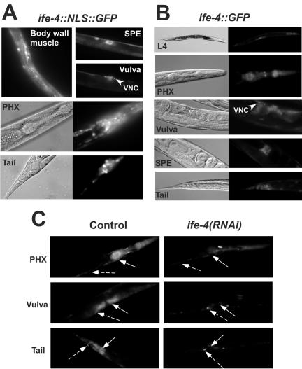
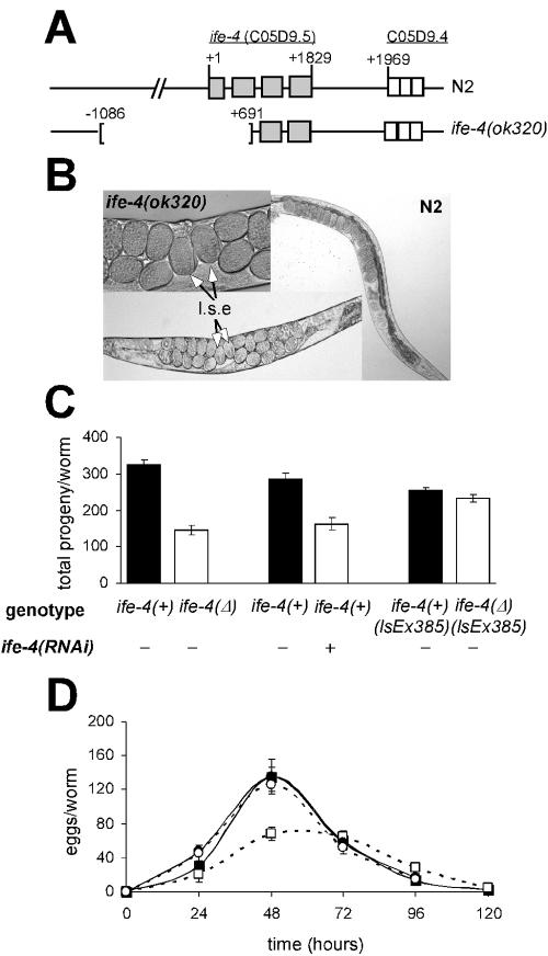
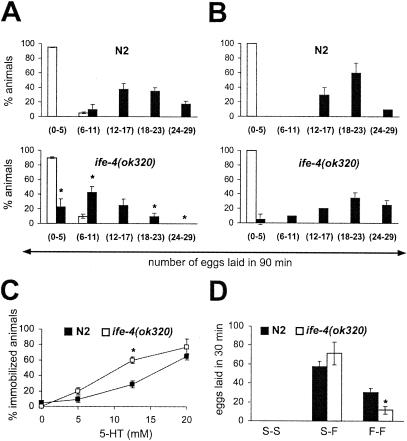
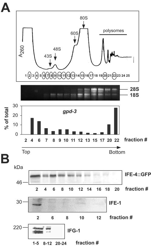
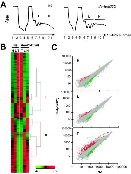
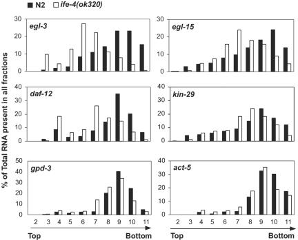
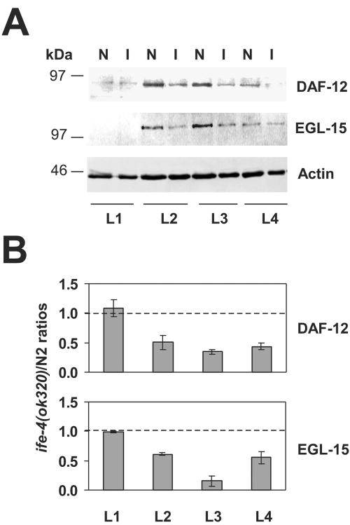
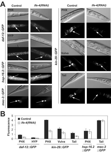
The mRNA cap-binding protein eukaryotic translation initiation factor 4E (eIF4E) participates in protein synthesis initiation, translational repression of specific mRNAs, and nucleocytoplasmic shuttling. Multiple isoforms of eIF4E are expressed in a variety of organisms, but their specific roles are poorly understood. We investigated one Caenorhabditis elegans isoform, IFE-4, which has homologues in plants and mammals. IFE-4::green fluorescent protein (GFP) was expressed in pharyngeal and tail neurons, body wall muscle, spermatheca, and vulva. Knockout of ife-4 by RNA interference (RNAi) or a null mutation produced a pleiotropic phenotype that included egg-laying defects. Sedimentation analysis demonstrated that IFE-4, but not IFE-1, was present in 48S initiation complexes, indicating that it participates in protein synthesis initiation. mRNAs affected by ife-4 knockout were determined by DNA microarray analysis of polysomal distribution. Polysome shifts, in the absence of total mRNA changes, were observed for only 33 of the 18,967 C. elegans mRNAs tested, of which a disproportionate number were related to egg laying and were expressed in neurons and/or muscle. Translational regulation was confirmed by reduced levels of DAF-12, EGL-15, and KIN-29. The functions of these proteins can explain some phenotypes observed in ife-4 knockout mutants. These results indicate that translation of a limited subset of mRNAs is dependent on a specific isoform of eIF4E.


Figures

FIG. 1.
FIG. 1.
Expression of ife-4::GFP in specific tissues of C. elegans (A and B) and reduction of fluorescence by ife-4(RNAi) (C). Transgenic strains were produced by microinjection of an ife-4 genomic fragment containing the complete coding region and ∼1,500 nt upstream fused to the 5′ end of DNA encoding GFP either as a single array with NLS [ife-4::NLS::GFP(lsEx296)] (A) or a complex array without NLS [ife-4::GFP(lsEx385)] (B). Fluorescence is seen in muscle and neurons from pharynx (PHX), neuronal cell bodies, the ventral nerve cord (VNC), vulval muscle and neurons, body wall muscle, and spermatheca (SPE). (C) RNAi was initiated for L4 ife-4::GFP(lsEx385) animals by feeding with bacteria expressing the dsRNA of ife-4 (right). Control animals were fed with bacteria containing empty vector (left). The offspring were compared for fluorescence at L4 after one generation. Solid and broken arrows indicate regions where fluorescence is decreased or not decreased by ife-4(RNAi), respectively.
FIG. 2.
FIG. 2.
Egl phenotype of the ife-4 homozygous deletion mutant. (A) Structure of the wild-type and truncated C. elegans ife-4 gene on the X chromosome. Light gray boxes indicate exons. For ife-4(ok320) animals, 1,776 bp were deleted. (B) Egg retention with ife-4(ok320) animals compared to that with N2 animals. Late-stage embryos (l.s.e) are shown. (C) The progeny laid during the reproductive life is reduced for ife-4(ok320) animals [ife-4(Δ)] and by ife-4(RNAi). Animals carrying the ife-4::GFP extrachromosomal transgene in an ife-4(ok320) background [ife-4(Δ) lsEx385] recover the normal number of progeny. (D) Egg laying for ife-4(Δ) (□) animals is reduced from that for N2 (▪) animals. Time after L4 is shown. ife-4(Δ) lsEx385 (○) animals had an egg-laying pattern similar to that of ife-4(+) lsEx385 (•) animals. The circles overlap in the graph. The results are means from five independent experiments.
FIG. 3.
FIG. 3.
Characterization of the Egl phenotype of ife-4(ok320) animals by serotonin (5-HT) and food assays. (A) ife-4(ok320) worms lay fewer eggs in response to 5-HT stimulation than N2 worms. The number of laid eggs was measured after 90 min of incubation in M9 (□) or 12.5 mM 5-HT (▪) for 20 animals per strain in three independent experiments. (B) The ife-4(ok320) egg-laying response to 5-HT increases after starvation. Conditions were the same as for panel A except that worms were deprived of food for 2 h previous to the 5-HT assay. (C) ife-4(ok320) animals are hypersensitive to 5-HT-induced locomotion arrest. Dose-response curves were generated in three independent experiments with 10 animals per strain per 5-HT concentration. The animals were scored for movement after 5 min in the presence of 5-HT. (D) Egg laying is impaired in fed but not starved ife-4(ok320) animals. The number of laid eggs was scored for 10 animals either starved (S-S), starved for 2 h then placed on food (S-F), or always on food (F-F). The experiment was performed in duplicate four independent times. Significant differences (P < 0.05) between ife-4(ok320) and N2 animals are indicated by an asterisk.
FIG. 4.
FIG. 4.
IFE-4 distribution in mRNPs and initiation complexes. Extracts from ife-4::GFP(lsEx385) worms were subjected to sedimentation on 10 to 30% sucrose gradients, and 0.5-ml fractions were collected. (A) The A260 profile (top) is shown together with the fractions analyzed for rRNA and gpd-3 mRNA distribution (circled). RNA was extracted as described in Materials and Methods and either subjected to electrophoresis on 1% agarose gels followed by ethidium bromide staining (middle) or assayed for gpd-3 mRNA by real-time PCR (bottom). Arrows indicate ribosomal subunits, initiation complexes, monosomes, and polysomes. (B) Proteins were precipitated with trichloroacetic acid, subjected to SDS-PAGE on 10% gels, and detected by Western blotting with primary antibody against GFP, IFE-1, or IFG-1. This experiment was performed three times with similar results.
FIG. 5.
FIG. 5.
Polysomal distributions of mRNAs in ife-4(ok320) and N2 animals. (A) Polysomal profiles (A260) of sucrose gradient fractions, subsequently pooled into H and L fractions. RNA was prepared from these fractions, as was total RNA (T) from whole worms. (B) Hierarchical clustering (http://www.cs.umd.edu/hcil/hce) of mRNAs judged present by Affymetrix GeneChip Array analysis in at least one sample (H, L, or T). Clustering was performed with signals normalized by the mean expression signal on the array and averaged over three independent experiments. Group I, mRNAs underrepresented in polysomes; Group II, mRNAs overrepresented in polysomes. (C) Scatter plots for the same signals in H, L, and T from ife-4(ok320) animals (y axis) versus N2 animals (x axis). Plots were made with the Genesifter.Net microarray data analysis system (VizV Labs LLC, Seattle, Wash.). Statistically significant (p < 0.05) differences of ≥2-fold are shown in color. Grey, no change; red, increased in ife-4(ok320) animals; green, decreased in ife-4(ok320) animals.
FIG. 6.
FIG. 6.
Polysomal shifts for selected mRNAs from ife-4(ok320) (□) versus N2 (▪) animals. Some of the mRNAs shown in Table 1 that were changed in polysomes (either H, L, or both) but not in T RNA (egl-3, egl-15, daf-12, or kin-29) were further analyzed over the entire polysomal gradient by real-time PCR. gpd-3 and actin (act-5) genes were selected as control mRNAs whose distribution did not change in H, L, or T fractions.
FIG. 7.
FIG. 7.
DAF-12 and EGL-15 protein levels are reduced in ife-4(ok320) animals. (A) Expression of DAF-12 and EGL-15 was detected by Western blotting at the indicated developmental stages (L1 to L4) for N2 (N) and ife-4(ok320) (I) animals. Proteins (50 μg) were resolved by SDS-PAGE on 10% gels. (B) DAF-12 and EGL-15 protein levels were normalized with the actin signal, and the ife-4(ok320)/N2 ratios are graphically represented, with 1.0 shown by the dashed line. The results represent the average for three independent worm populations and immunoblots.
FIG. 8.
FIG. 8.
ife-4(RNAi) feeding reduces the expression of daf-12A::GFP and kin-29::GFP but not of hsp-16.2::GFP or mec-3::GFP in specific tissues. (A) RNAi feeding was performed as described in the legend to Fig. 1. Representative Nomarski (upper) and fluorescence (lower) images are shown for each condition in different tissues. Arrows indicate regions measured for fluorescence quantification. At least five different animals were measured for each condition, and the results were averaged. PHX, pharynx; HYP, hypodermis. (B) The graphical representation indicates the maximum fluorescence for the different strains in various tissues.

References

    1. Amiri, A., B. D. Keiper, I. Kawasaki, Y. Fan, Y. Kohara, R. E. Rhoads, and S. Strome. 2001. An isoform of eIF4E is a component of germ granules and is required for spermatogenesis in C. elegans. Development 128:3899-3912. - PMC - PubMed
    1. Antebi, A., W.-H. Yeh, D. Tait, E. M. Hedgecock, and D. L. Riddle. 2000. daf-12 encodes a nuclear receptor that regulates the dauer diapause and developmental age in C elegans. Genes Dev. 14:1512-1527. - PMC - PubMed
    1. Bradley, C. A., J. C. Padovan, T. L. Thompson, C. A. Benoit, B. T. Chait, and R. E. Rhoads. 2002. Mass spectrometric analysis of the N-terminus of translational initiation factor eIF4G-1 reveals novel isoforms. J. Biol. Chem. 277:12559-12571. - PubMed
    1. Brenner, S. 1974. The genetics of Caenorhabditis elegans. Genetics 77:71-94. - PMC - PubMed
    1. Browning, K. S., C. Webster, J. K. M. Roberts, and J. M. Ravel. 1992. Identification of an isozyme form of protein synthesis initiation factor 4F in plants. J. Biol. Chem. 267:10096-10100. - PubMed

Publication types

MeSH terms

LinkOut - more resources