. 2005 Jan;25(1):162-71.
doi: 10.1128/MCB.25.1.162-171.2005.

BTB protein Keap1 targets antioxidant transcription factor Nrf2 for ubiquitination by the Cullin 3-Roc1 ligase

Affiliations

BTB protein Keap1 targets antioxidant transcription factor Nrf2 for ubiquitination by the Cullin 3-Roc1 ligase

Manabu Furukawa et al. Mol Cell Biol. 2005 Jan.

Abstract

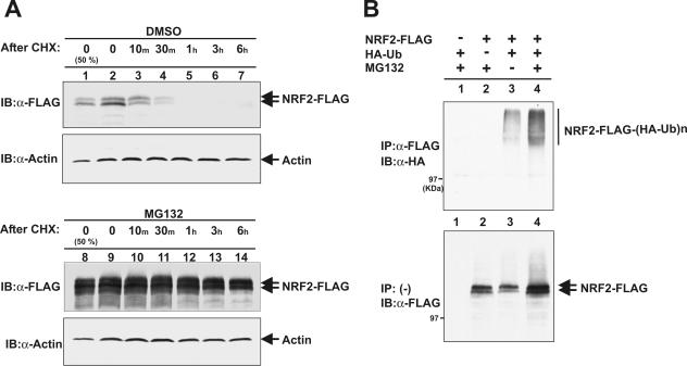
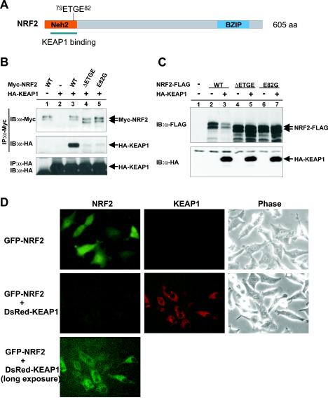
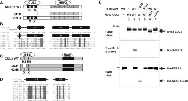
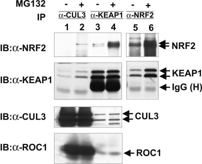
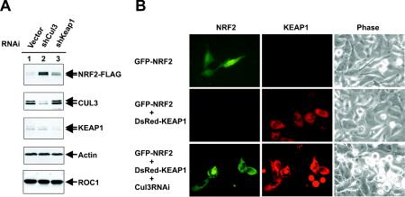
The concentrations and functions of many eukaryotic proteins are regulated by the ubiquitin pathway, which consists of ubiquitin activation (E1), conjugation (E2), and ligation (E3). Cullins are a family of evolutionarily conserved proteins that assemble by far the largest family of E3 ligase complexes. Cullins, via a conserved C-terminal domain, bind with the RING finger protein Roc1 to recruit the catalytic function of E2. Via a distinct N-terminal domain, individual cullins bind to a protein motif present in multiple proteins to recruit specific substrates. Cullin 3 (Cul3), but not other cullins, binds directly with BTB domains to constitute a potentially large number of BTB-CUL3-ROC1 E3 ubiquitin ligases. Here we report that the human BTB-Kelch protein Keap1, a negative regulator of the antioxidative transcription factor Nrf2, binds to CUL3 and Nrf2 via its BTB and Kelch domains, respectively. The KEAP1-CUL3-ROC1 complex promoted NRF2 ubiquitination in vitro and knocking down Keap1 or CUL3 by short interfering RNA resulted in NRF2 protein accumulation in vivo. We suggest that Keap1 negatively regulates Nrf2 function in part by targeting Nrf2 for ubiquitination by the CUL3-ROC1 ligase and subsequent degradation by the proteasome. Blocking NRF2 degradation in cells expressing both KEAP1 and NRF2 by either inhibiting the proteasome activity or knocking down Cul3, resulted in NRF2 accumulation in the cytoplasm. These results may reconcile previously observed cytoplasmic sequestration of NRF2 by KEAP1 and suggest a possible regulatory step between KEAP1-NRF2 binding and NRF2 degradation.


Figures

FIG. 1.
FIG. 1.
NRF2 is rapidly ubiquitinated and degraded by proteasomes. (A) 293T cells were transfected with the plasmid expressing FLAG-tagged NRF2 (NRF2-FLAG). Twenty-four hours after transfection, cells were treated first with MG132 (25 μM) or dimethyl sulfoxide (DMSO) for 5 h and then with cycloheximide (CHX) (75 μg/ml) for periods of time ranging from 10 min to 6 h. Cells were lysed, and 50 μg (lane 1) or 100 μg (lanes 2 to 7) of total lysates were resolved by SDS-PAGE, followed by immunoblotting (IB) with anti-FLAG (α-FLAG) or antiactin (α-Actin) antibodies. (B) 293T cells were transfected with the indicated plasmids expressing FLAG-tagged NRF2 and HA-tagged ubiquitin (HA-Ub). Twenty hours after transfection, cells were treated with MG132 (25 μM) for 2 h prior to cell lysis. Cells were lysed in a SDS lysis buffer and boiled for 15 min. Lysates were then diluted with NP-40 lysis buffer and immunoprecipitated (IP) with anti-FLAG antibody. The washed immunoprecipitates and 50 μg of lysates were resolved by SDS-PAGE, followed by immunoblotting with anti-HA and anti-FLAG antibodies, respectively.
FIG. 2.
FIG. 2.
KEAP1 promotes NRF2 degradation. (A) Schematic illustration of NRF2 protein and mutations characterized in this study. BZIP, basic region leucine zipper; aa, amino acids. (B) 293T cells were cotransfected with the plasmids expressing HA-tagged KEAP1 (HA-KEAP1) and Myc-tagged NRF2 (Myc-NRF2) (wild-type [WT] or mutant NRF2). Twenty hours after transfection, cells were treated with MG132 (25 μM) for 4 h prior to lysis, and the NRF2-KEAP1 association was examined by IP-Western. IB, immunoblotting; α-Myc, anti-Myc antibody; α-HA, anti-HA antibody. (C) HeLa cells were cotransfected with the plasmids expressing HA-tagged KEAP1 and FLAG-tagged wild-type or mutant NRF2. Twenty-four hours after transfection, cells were lysed, and lysates (0.1 mg) were resolved by SDS-PAGE, followed by immunoblotting (IB) with anti-FLAG (α-FLAG) or anti-HA antibodies. (D) GFP-tagged NRF2 was transfected to HeLa cells either alone or with the plasmid expressing DsRed-tagged KEAP1. Twenty-four hours after transfection, the levels of expression of GFP-NRF2 and DsRed-KEAP1 were examined by fluorescence or phase-contrast microscopy.
FIG. 3.
FIG. 3.
BTB domain in Keap1 binds to the N terminus of CUL3. (A) Schematic illustration of wild-type (WT) and mutant Keap1. (B) Conserved α/β-structure in BTB/POZ fold of SKP1, PLZF, and Keap1. Identical and conserved residues are indicated by dark gray and light gray shading, respectively. Residues in the SKP1 making contact with CUL1 are marked by asterisks. The mutated residues in the β-sheet S3 and α-helix H4 (KEAP1S3H4 mutant) are shown at the bottom. (C) Schematic figures of wild-type CUL3, ΔN41 (CUL3 in which the N-terminal 41 residues were deleted), and helix 2 helix 5 mutant (H2H5) CUL3. (D) Sequence comparison of helix 2 (H2) and helix 5 (H5) of CUL1 and CUL3. Residues in the H2 and H5 helices of CUL1 that make contact with SKP1 are marked by asterisks. Residues conserved in CUL3 are marked by shading. The residues mutated in the CUL3H2H5 mutant are shown at the bottom. (E) 293T cells were cotransfected with the indicated plasmids expressing Myc-tagged CUL3 (Myc3-CUL3) or HA-tagged KEAP1 (HA-KEAP1) (wild-type [WT] or mutant CUL3 or KEAP1). Twenty-four hours after transfection, cells were lysed, and the CUL3-KEAP1 association was examined by IP-Western. IB, immunoblotting; α-Myc, anti-Myc antibody.
FIG. 4.
FIG. 4.
Keap1 binds to Nrf2 and CUL3 directly. (A) His-tagged NRF2 (His-NRF2) fusion protein was purified from bacteria and mixed with bacterially purified GST or GST-KEAP1 fusion proteins immobilized on the glutathione agarose beads. NRF2-KEAP1 binding was examined by immunoblotting (IB). α-NRF2, anti-NRF2 antibody; α-GST, anti-GST antibody. (B) His-CUL3N197 fusion protein was purified from bacteria and mixed with bacterially purified GST or GST-KEAP1 fusion proteins immobilized on the glutathione agarose beads. CUL3N197-KEAP1 binding was examined by immunoblotting (IB). α-CUL3, anti-CUL3 antibody.
FIG. 5.
FIG. 5.
Endogenous CUL3, KEAP1, and NRF2 form a complex. 293T cells were treated with MG132 or DMSO (−) for 4 h prior to lysis. Cell lysates were immunoprecipitated (IP) with antibodies to CUL3 (α-CUL3), KEAP1 (α-KEAP1), or NRF2 (α-NRF2). Washed immunocomplexes were resolved by SDS-PAGE, followed by immunoblotting (IB) with the indicated antibodies. IgG (H), immunoglobulin G (heavy chain).
FIG. 6.
FIG. 6.
KEAP1-CUL3-ROC1 complex ubiquitinates NRF2 in vitro. (A) KEAP1-CUL3-ROC1 complex. 293T cells were cotransfected with the plasmids expressing the indicated proteins, Myc-tagged CUL3 (Myc-CUL3), T7-tagged KEAP1 (T7-KEAP1), and HA-tagged ROC1 (HA-ROC1). Twenty-four hours after transfection, cells were lysed and immunoprecipitated (IP) with anti-Myc antibody (α-Myc). Total lysates (lane 1 and 2) and immunoprecipitates (lanes 3 and 4) were resolved by SDS-PAGE, followed by immunoblotting (IB) with the indicated antibodies. (B) In vitro ubiquitination of NRF2. KEAP1-CUL3-ROC1 complexes were prepared from triply transfected cells by immunoprecipitation using myc antibody and used as the source of E3 ligase. Bacterially expressed and purified FLAG-tagged NRF2 was incubated with KEAP1-CUL3-ROC1 complex, E1, E2, ubiquitin (Ub), and ATP, and reaction mixtures were resolved by SDS-PAGE and immunoblotted with the indicated antibodies.
FIG. 7.
FIG. 7.
NRF2 accumulated when Cul3 or Keap1 was silenced. (A) 293T cells were cotransfected with the plasmid expressing FLAG-tagged NRF2 and the indicated short hairpin RNA plasmids (shCul3 and shKeap1). Transfected cells were selected by puromycin (1 μg/ml). Ninety-six hours after transfection, selected cells were lysed, and 50-μg samples of total lysates were resolved by SDS-PAGE and immunoblotted with the indicated antibodies. RNAi, RNA interference. (B) The expression vector for GFP-NRF2 alone or for GFP-NRF2 and DsRed-Keap1 was cotransfected with CUL3 short hairpin RNA plasmid or control short hairpin RNA plasmid to HeLa cells. After puromycin selection, the expression and localization of GFP-NRF2 and DsRed-KEAP1 were examined by fluorescence or phase-contrast microscopy.

References

    1. Ahmad, K. F., C. K. Engel, and G. G. Prive. 1998. Crystal structure of the BTB domain from PLZF. Proc. Natl. Acad. Sci. USA 95:12123-12128. - PMC - PubMed
    1. Bai, C., P. Sen, K. Hofmann, L. Ma, M. Goebl, J. W. Harper, and S. J. Elledge. 1996. SKP1 connects cell cycle regulators to the ubiquitin proteolysis machinery through a novel motif, the F-box. Cell 86:263-274. - PubMed
    1. Balogun, E., M. Hoque, P. Gong, E. Killeen, C. J. Green, R. Foresti, J. Alam, and R. Motterlini. 2003. Curcumin activates the haem oxygenase-1 gene via regulation of Nrf2 and the antioxidant-responsive element. Biochem. J. 371:887-895. - PMC - PubMed
    1. Bloom, D. A., and A. K. Jaiswal. 2003. Phosphorylation of Nrf2 at Ser40 by protein kinase C in response to antioxidants leads to the release of Nrf2 from INrf2, but is not required for Nrf2 stabilization/accumulation in the nucleus and transcriptional activation of antioxidant response element-mediated NAD(P)H:quinone oxidoreductase-1 gene expression. J. Biol. Chem. 278:44675-44682. - PubMed
    1. Chen, A., K. Wu, S. Y. Fuchs, P. Tan, C. Gomez, and Z. Q. Pan. 2000. The conserved RING-H2 finger of ROC1 is required for ubiquitin ligation. J. Biol. Chem. 275:15432-15439. - PubMed

Publication types

MeSH terms