. 2005 Jan;25(1):197-205.
doi: 10.1128/MCB.25.1.197-205.2005.

Overcoming functional redundancy to elicit pachyonychia congenita-like nail lesions in transgenic mice

Affiliations

Overcoming functional redundancy to elicit pachyonychia congenita-like nail lesions in transgenic mice

Pauline Wong et al. Mol Cell Biol. 2005 Jan.

Abstract

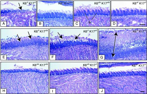
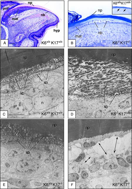
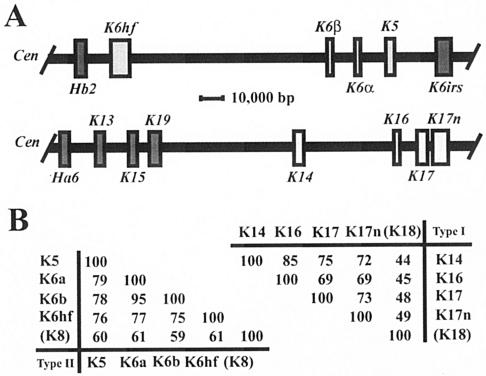
Mutations affecting the coding sequence of intermediate filament (IF) proteins account for >30 disorders, including numerous skin bullous diseases, myopathies, neuropathies, and even progeria. The manipulation of IF genes in mice has been widely successful for modeling key features of such clinically distinct disorders. A notable exception is pachyonychia congenita (PC), a disorder in which the nail and other epithelial appendages are profoundly aberrant. Most cases of PC are due to mutations in one of the following keratin-encoding genes: K6, K16, and K17. Yet null alleles obliterating the function of both K6 genes (K6alpha and K6beta) or the K17 gene, as well as the targeted expression of a dominant-negative K6alpha mutant, elicit only a subset of PC-specific epithelial lesions (excluding that of the nail in mice). We show that newborn mice null for K6alpha, K6beta, and K17 exhibit severe lysis restricted to the nail bed epithelium, where all three genes are robustly expressed, providing strong evidence that this region of the nail unit is initially targeted in PC. Our findings point to significant redundancy among the multiple keratins expressed in hair and nail, which can be related to the common ancestry, clustered organization, and sequence relatedness of specific keratin genes.


Figures

FIG. 1.
FIG. 1.
Earlier lysis of dorsal tongue epithelium in K6α/K6β−/− K17−/− null neonatal mice compared to other genotypes. Tongues from P0 (A to D), and P2 (E to J) mice were surgically removed and processed for routine histology. Micrographs shown are derived from H&E-stained preparations of longitudinally sectioned tongue tissue (rostrocaudal axis). The mouse genotype is indicated in the upper right corner. (A to D) Lysis and destruction of the dorsal tongue epithelium can be seen as early as P0 in K6α/K6β−/− K17−/− tongue (see arrow in panel A). At that time, K6α/K6β−/− K17+/+ mice show comparatively mild lesions in the same region (B) whereas wild-type mice (C) and even K6α/K6β+/− K17−/− mice (D) show normal histology with intact filiform papillae (fp). Bar, 100 μm (A to D). (E to J) At P2, obvious signs of oral lesions can be detected in K6α/K6β−/− K17+/+ mice (see arrows in panel E) as reported previously (54). These lesions are significantly more severe and inflamed in K6α/K6β−/− K17+/− (see arrows in panel F) and especially in K6α/K6β−/− K17−/− mice (see arrow in panel G). In contrast, the dorsal epithelial tongue appears normal in K6α/K6β+/+ K17+/+ (wild-type) mice (H), as expected, and in K6α/K6β+/+ K17−/− (I) and K6α/K6β+/− K17−/− (J) mice as well. Bar, 200 μm (E to J).
FIG.2.
FIG.2.
Early lysis of the nail bed epithelium in K6α/K6β−/− K17−/− null newborn mice but not in other genotypes. Paws or digits from P0 mice (newborns) were surgically removed and processed for routine histology. Micrographs shown are derived from an H&E-stained section of paraffin-embedded tissue (A), toluidine-blue stained semithin sections (∼0.5 μm thick) prepared from epoxy-embedded tissues (B), and uranyl acetate- and lead citrate-counterstained stained ultrathin sections (∼50 to 70 nm thick) prepared from epoxy-embedded tissue (C to F). In all cases, the sectioning plane is along the anterior-posterior axis and the mouse genotype is indicated in the lower right corner. (A) Section of wild-type nail tissue showed for orientation. The proximal nail fold (pnf), matrix (mat), nail bed (nb), nail plate (np), and hyponychium (hyp) compartments are depicted. Bar, 300 μm. (B) Semithin section of epoxy-embedded K6α/K6β−/− K17−/− nail unit, depicting the nail bed area. There is striking lysis of the nail bed (nb) epithelium, while the matrix (mat) and nail plate (np) appear not affected. The inset shows an identical preparation from a wild-type mouse, which does not show any nail bed (nb) alteration. (C to F) Electron microscopy data taken from a smaller subset of the same general area (nail bed). In contrast to K6α/K6β+/+ K17+/+ (C), K6α/K6β−/− K17+/+ (D), and K6α/K6β+/+ K17+/+ (E) mice, the nail bed of K6α/K6β−/− K17−/− mice (F) shows obvious ballooning and lysis in the area immediate above the basal layer (see arrows). Likewise, the two to three layers of cells with dense keratin bundles (kb in panels C to E) are missing from the K6α/K6β−/− K17−/− sample (F). In contrast, the nail plate (np) and matrix appear normal in these mice. n, nucleus. Bar, 5 μm (C to F).
FIG. 3.
FIG. 3.
Analysis of the levels and distribution of specific keratin antigens in newborn skin keratinocytes in primary culture. (A) Western immunoblot analysis. The genotype is indicated on top, and the specificity of the antiserum used is given at the left of each blot. For each genotype, primary keratinocytes from three different mice were pooled and used. A total of 2 μg of proteins was loaded per lane. Bound primary antibodies were detected by chemiluminescence. Actin was used as a loading control (55). (B) Keratinocytes cultures from various genotypes, indicated at top of each pair of micrographs, were fixed and immunostained with antibodies directed towards K5 (left column) and K16 (right column). Wild-type keratinocytes exhibit pan-cytoplasmic IF networks (arrowheads). A subset of keratinocytes, identified with arrows, show altered and even collapsed keratin networks for the K6α/K6β−/− K17+/− and K6α/K6β−/− K17−/− genotypes. The number of cells affected and the severity of the disruption are clearly higher in the latter. There is no significant difference in the results obtained when staining for either K5 or K16 antigen, consistent with the known ability of several keratins to copolymerize into IFs. Bar, 50 μm.
FIG. 4.
FIG. 4.
Clustering of related keratin genes on mouse chromosomes 15 and 11. (A) Organization of subsets of type II and type I keratin genes on mouse chromosomes 15 (top) and 11 (bottom), respectively. In both instances the centromere (Cen) is located on the left side. All keratin genes located in these two clusters, including the ones shown here, display the same transcriptional orientation towards the centromere. All type II keratin genes exhibit nine introns, whereas all type I genes exhibit eight introns, located in each case at identical positions within the coding sequence (data not shown; see reference 16). The genes of interest to this study are displayed in light-shaded boxes. This information was adapted from Wang et al. (48) and Tong and Coulombe (46). (B) Pairwise comparisons of the primary structures for type II (left) and type I (right) keratins expressed in the nail bed epithelium. Percent identity scores, as determined by the maximum matching routine in DNAsis version 3.5 software (Hitachi, Tokyo, Japan), are shown for the sequences encoding the nonhelical head and central rod domain of these keratins. K8 and K18, which represent the major keratin pair expressed in simple epithelia, have been included for reference purposes. The physical proximity, identical substructures, identical orientations of transcription, and obvious sequence relatedness of the type II keratin genes K5, K6α, K6β, and K6hf as well as the type I genes K14, K16, K17, and K17n strongly suggest that they were each generated through successive duplications from a common ancestral gene. The information needed to construct this figure is derived from the mouse genome browser available at http://www.ensembl.org/. The same principles apply to the human orthologs for these genes (4).

References

    1. Arin, M. J., and D. R. Roop. 2001. Disease model: heritable skin blistering. Trends Mol. Med. 7:422-424. - PubMed
    1. Bernot, K., K. McGowan, and P. A. Coulombe. 2002. Keratin 16 expression defines a subset of epithelial cells during skin morphogenesis and the hair cycle. J. Investig. Dermatol. 119:1137-1149. - PubMed
    1. Blumenberg, M. 1988. Concerted gene duplications in the two keratin gene families. J. Mol. Evol. 27:203-211. - PubMed
    1. Coulombe, P. A., and K. M. Bernot. 2004. Keratins and the skin, p. 497-504. In M. D. Lane, and W. Lennarz, (ed.), Encyclopedia of biological chemistry. Elsevier, Oxford, United Kingdom.
    1. Coulombe, P. A., L. Ma, S. Yamada, and M. Wawersik. 2001. Intermediate filaments at a glance. J. Cell Sci. 114:4345-4347. - PubMed

Publication types

MeSH terms