. 2005 Jan;25(1):250-63.
doi: 10.1128/MCB.25.1.250-263.2005.

A novel intronic cis element, ISE/ISS-3, regulates rat fibroblast growth factor receptor 2 splicing through activation of an upstream exon and repression of a downstream exon containing a noncanonical branch point sequence

Affiliations

A novel intronic cis element, ISE/ISS-3, regulates rat fibroblast growth factor receptor 2 splicing through activation of an upstream exon and repression of a downstream exon containing a noncanonical branch point sequence

Ruben H Hovhannisyan et al. Mol Cell Biol. 2005 Jan.

Abstract

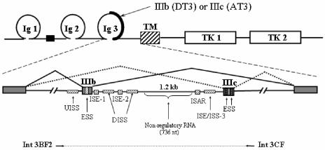
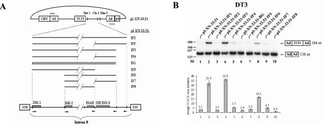
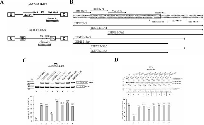
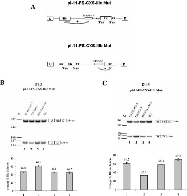
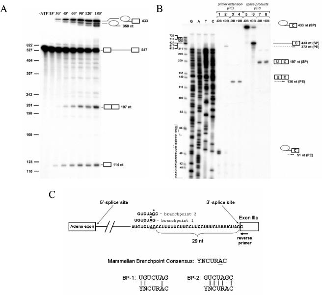
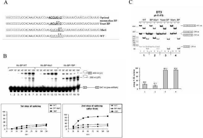
Mutually exclusive splicing of fibroblast growth factor receptor 2 (FGFR2) exons IIIb and IIIc yields two receptor isoforms, FGFR2-IIIb and -IIIc, with distinctly different ligand binding properties. Several RNA cis elements in the intron (intron 8) separating these exons have been described that are required for splicing regulation. Using a heterologous splicing reporter, we have identified a new regulatory element in this intron that confers cell-type-specific inclusion of an unrelated exon that mirrors its ability to promote cell-type-specific inclusion of exon IIIb. This element promoted inclusion of exon IIIb while at the same time silencing exon IIIc inclusion in cells expressing FGFR2-IIIb; hence, we have termed this element ISE/ISS-3 (for "intronic splicing enhancer-intronic splicing silencer 3"). Silencing of exon IIIc splicing by ISE/ISS-3 was shown to require a branch point sequence (BPS) using G as the primary branch nucleotide. Replacing a consensus BPS with A as the primary branch nucleotide resulted in constitutive splicing of exon IIIc. Our results suggest that the branch point sequence constitutes an important component that can contribute to the efficiency of exon definition of alternatively spliced cassette exons. Noncanonical branch points may thus facilitate cell-type-specific silencing of regulated exons by flanking cis elements.


Figures

FIG. 1.
FIG. 1.
Schematic representation of alternatively spliced variants of FGFR2 and RNA cis elements shown to regulate splicing. At the top is a protein domain map showing the region encoded by exons IIIb or IIIc. At the bottom is a map of the pre-mRNA with mutually exclusive pathways leading to production of FGFR2-IIIb in DT3 cells or FGFR2-IIIc in AT3 cells. TM, transmembrane domain; TK, tyrosine kinase domains. Shaded boxes represent exons, and solid lines represent introns. Hatched boxes represent intronic and exonic cis elements (enhancers and silencers). Note that human sequences highly similar to ISE-1, ISE-2, and ISAR have been referred to as IAS1, IAS2, and IAS3, respectively, and can be considered to be functionally equivalent. The horizontal arrows and dashed line underneath the pre-mRNA represent the FGFR2 genomic sequence that was amplified by PCR and positioned in the intron of pI-11 to generate the full-length FGFR2 minigenes as described in Results.
FIG. 2.
FIG. 2.
Intron 8 sequences from FGFR2 can activate splicing of an upstream heterologous troponin I (TNI) exon in DT3 cells but not in AT3 cells. (A) Schematic presentation of pI-XN-33.51 series plasmids and intron 8 elements from FGFR2 that are positioned downstream of exon 33.51. Ad, adenoviral exons; CMV, cytomegalovirus exons. Solid lines represent intronic fragments (IF) that were inserted downstream of the TNI exon 33.51. Diagonal lines indicate deleted sequence, and the cross-hatched box represents a β-globin intron sequence. Outlined arrows indicate primers used for RT-PCR analysis. At the bottom is a representation of intron 8 to illustrate the derivation of the intron fragments. Open boxes indicate the exons, and solid arrows indicate positions of PCR primers used to generate the intron fragments. (B) Intron 8 sequences from FGFR2 can activate splicing of a heterologous exon in DT3 cells. At the top is shown a representation of a single experiment using RT-PCR of RNA from DT3 cells stably transfected with these minigenes, and at the bottom is shown graphical results representing the average of at least three independent experiments (this same format applies to most subsequent transfection results). RT-PCR products with inclusion or skipping of the cTNI exon are indicated at the right. M, molecular weight markers.
FIG. 3.
FIG. 3.
An 85-nt sequence, ISE/ISS-3Δ6, is sufficient to restore splicing regulation by sequences downstream of ISAR. (A) Schematic representation of the minigene constructs used to study the influence of ISE/ISS-3 deletions and mutations on splicing of exon 33.51 splicing in the heterologous minigenes (top) or on exon IIIb and IIIc splicing in the full-length FGFR2 minigenes (bottom). (B) Sequence of the ISE/ISS-3 RNA cis element. Deletions from the 5′ or 3′ end were obtained by PCR of the corresponding DNA with primer positions indicated by arrows. The resulting DNA sequences were inserted between ClaI and XhoI sites in the pI-XN-33.51-IF5 and pI-11-FS-CXS plasmids. The boxed sequence indicates the minimal 78-nt sequence that is sufficient for full ISE/ISS-3 function. Underlined is a sequence that, when mutated to the sequence shown above (Mut), results in loss of exon 33.51 inclusion and a switch from exon IIIb to IIIc in full-length minigenes. (C) Results of RT-PCR of RNA prepared from DT3 cells stably transfected by the minigenes containing the truncated sequences downstream of the heterologous cTNI exon 33.51. (D) Results of RT-PCR and restriction analysis to determine the percentage of exon IIIb inclusion using full-length FGFR2 minigenes in DT3 cells. When undigested (U) RT-PCR products are analyzed, three bands are observed: a 431-bp product corresponding to a spliced product containing both exon IIIb and IIIc (double inclusion), a 138-bp band corresponding to spliced products in which both exon IIIb and IIIc are skipped, and 286- or 283-bp products corresponding to products containing either exon IIIb or exon IIIc (single inclusion), respectively, that cannot be easily distinguished from each other by size. To distinguish exon IIIb from exon IIIc, the RT-PCR products are digested with AvaI (A) or HincII (H), which recognize restriction sites present only in exon IIIb- or exon IIIc-containing products, respectively. We calculated the percentage of the single-inclusion products that contain exon IIIb versus exon IIIc, and the results are presented graphically at the bottom. U, undigested RT-PCR products; A, products digested with AvaI; H, products digested with HincII. M, pBR322/MspI molecular weight markers. At the right-hand side, spliced products represented by each band are shown. U and D indicate the upstream and downstream adenoviral exons; B and C, exons IIIb and IIIc, respectively.
FIG. 4.
FIG. 4.
ISE/ISS-3 enhances splicing of exon IIIb and represses splicing of exon IIIc. (A) Schematic representation of the constructs showing splice site mutations in which GG and CT replace the AG and GU dinucleotides, respectively. (B) Results of RT-PCR for pI-11-FS-CXS-IIIc Mut minigene series stable transfected in DT3 cells. Results in which no sequence is inserted to replace ISE/ISS-3, ISE/ISS-3, ISE/ISS-3 Mut, or the unrelated BG sequence are shown. (C) Similar results showing the effect of ISE/ISS-3 on exon IIIc splicing using the pI-11-FS-CXS-IIIb Mut minigenes. ss, splice site; U, D, and M, as defined in the legend to Fig. 3.
FIG. 5.
FIG. 5.
Branch point mapping for exon IIIc. (A) A time course of in vitro splicing of a pre-mRNA containing an upstream adenoviral exon followed by FGFR2 intron 8 sequences and exon IIIc. From top to bottom, icons at the right indicate the lariat intermediate, lariat product, pre-mRNA, spliced product, and free 5′ exon. (B) Branch point determination by primer extension. At the far right-hand side, icons present splicing products. Arrows indicate the primer used for sequencing and primer extension, and dotted lines show products of the primer extension. Samples in lines 1 to 4 were primer extended (PE) before loading the gel; samples in lines 5 to 8 represent gel-purified products from the spliced reaction. +DB, debranched; −DB, not debranched. At far left are ΦX174/HinfI molecular weight markers followed by a sequencing ladder obtained using the same primer that was used for primer extension. Sequence of the minus strand and the positions at which primer extension is blocked are indicated by stars. (C) Schematic presentation of branch point sequences identified. On the bottom, the degree of matching of the branch point sequences to the degenerate mammalian BPS consensus is shown. Adeno, adenovirus.
FIG. 6.
FIG. 6.
Replacement of the weak exon IIIc branch point sequence with consensus branch points rescues the second step of splicing in vitro and leads to constitutive inclusion of exon IIIc in DT3 cells in vivo. (A) Sequences showing alterations in the branch point region upstream of exon IIIc. (B) A time course of in vitro splicing reactions with pre-mRNAs containing the wild type (WT), A-to-G-mutated branch point (Mut1), and yeast branch point sequence in HeLa nuclear extract. Above each gel the duration of incubation in HeLa cell nuclear extracts is indicated. At the right, icons indicate (from top to bottom) lariat intermediate, lariat product, and pre-mRNA. The lower graphs show the efficiency of the first and second steps of splicing of exon IIIc. Calculation of the first-step efficiency represents the percentage of pre-mRNA processed to lariat intermediate (LI) or lariat product (LP) (LI + LP/LI + LP + pre-mRNA). The efficiency of the second step (after step 1) represents the percentage of products undergoing the first step that also complete the second step (LI/LI + LP). (C) Results of RT-PCR of RNA prepared from DT3 cells stably transfected by the minigenes containing the indicated branch point sequences. Lane and band designations are the same as those described in the legend for Fig. 3D. In the graph, the percentage of exon IIIc inclusion in single-inclusion products is indicated compared to that of exon IIIb inclusion.
FIG. 7.
FIG. 7.
Diagram demonstrating the sequences present between ISAR and ISE/ISS-3. GCAUG elements are boxed where present, with the unaltered wild-type (WT) sequence junction for pI-11-FS and pI-XN-33.51-IF3 showing two overlapping copies. Underlined sequences represent those encoded by ClaI or XhoI sites inserted to facilitate study of ISE/ISS-3. pI-11-FS-CXS and pI-XN-33.51-IF5 indicate the sequences in which no ISE/ISS-3 element was inserted into the ClaI and XhoI sites.
FIG. 8.
FIG. 8.
Schematic illustrating two models in which the noncanonical branch point facilitates inhibition of exon IIIc inclusion in DT3 cells. In model A, a factor(s) associated with ISE/ISS-3 (and, potentially, other factors) is able to prevent the first step of splicing by preventing recognition of the exon IIIc weak branch point sequence during commitment complex (CC or E complex) formation or A complex assembly. As a result, the branch point associated with exon 9 forms a lariat structure with the 5′ end of intron 8 and exon IIIc is sequestered in the lariat intermediate. Ligation of exon IIIb (3B) to exon 9 ensues in the second step. In model B, the first step of splicing can occur using the weak exon IIIc branch nucleotide(s) to generate the lariat intermediate shown. However, ISE/ISS-3-associated factors (and possibly others as well) are able to prevent the second step of splicing using the 3′ splice site of exon IIIc. Thus, ligation of exon IIIb to exon 9 occurs instead. Note that only the branch G nucleotide upstream of exon IIIc is shown for clarity. Also omitted are other intron 8 elements that may also contribute to exon IIIc silencing and/or exon IIIb activation.

References

    1. Abovich, N., and M. Rosbash. 1997. Cross-intron bridging interactions in the yeast commitment complex are conserved in mammals. Cell 89:403-412. - PubMed
    1. Adema, G. J., R. A. Bovenberg, H. S. Jansz, and P. D. Baas. 1988. Unusual branch point selection involved in splicing of the alternatively processed calcitonin/CGRP-I pre-mRNA. Nucleic Acids Res. 16:9513-9526. - PMC - PubMed
    1. Adema, G. J., K. L. van Hulst, and P. D. Baas. 1990. Uridine branch acceptor is a cis-acting element involved in regulation of the alternative processing of calcitonin/CGRP-l pre-mRNA. Nucleic Acids Res. 18:5365-5373. - PMC - PubMed
    1. Baraniak, A. P., E. L. Lasda, E. J. Wagner, and M. A. Garcia-Blanco. 2003. A stem structure in fibroblast growth factor receptor 2 transcripts mediates cell-type-specific splicing by approximating intronic control elements. Mol. Cell. Biol. 23:9327-9337. - PMC - PubMed
    1. Berget, S. M. 1995. Exon recognition in vertebrate splicing. J. Biol. Chem. 270:2411-2414. - PubMed

Publication types

MeSH terms

LinkOut - more resources