. 2004 Dec 15;24(50):11236-43.
doi: 10.1523/JNEUROSCI.3724-04.2004.

Subcortical modulation of attention counters change blindness

Affiliations

Subcortical modulation of attention counters change blindness

James Cavanaugh et al. J Neurosci. .

Abstract

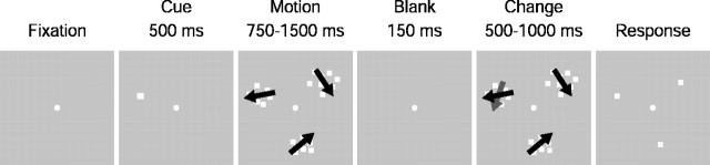
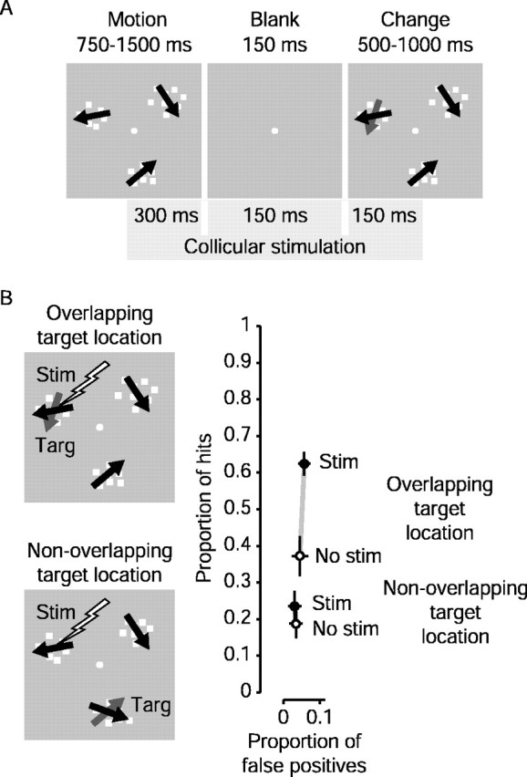
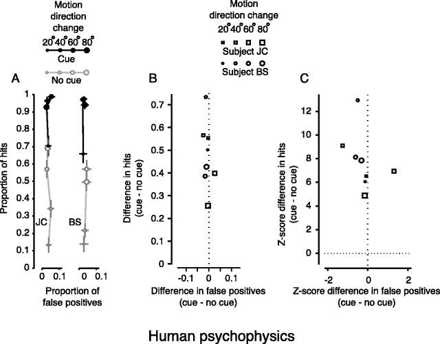
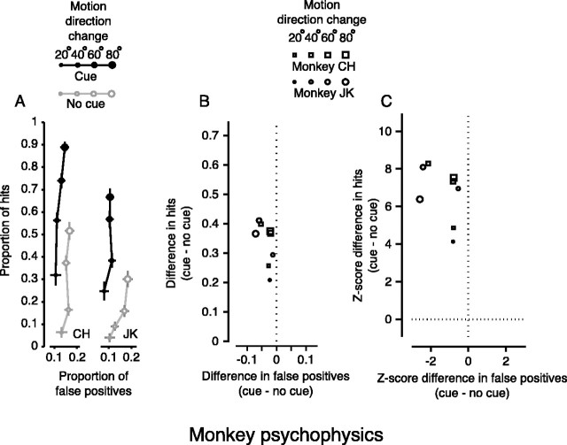
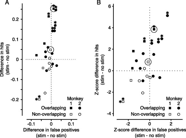
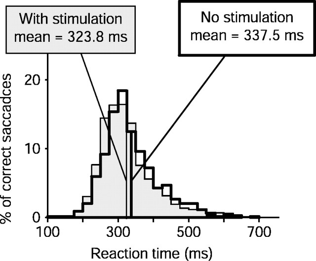
Change blindness is the failure to see large changes in a visual scene that occur simultaneously with a global visual transient. Such visual transients might be brief blanks between visual scenes or the blurs caused by rapid or saccadic eye movements between successive fixations. Shifting attention to the site of the change counters this "blindness" by improving change detection and reaction time. We developed a change blindness paradigm for visual motion and then showed that presenting an attentional cue diminished the blindness in both humans and old world monkeys. We then replaced the visual cue with weak electrical stimulation of an area in the monkey's brainstem, the superior colliculus, to see if activation at such a late stage in the eye movement control system contributes to the attentional shift that counters change blindness. With this stimulation, monkeys more easily detected changes and had shorter reaction times, both characteristics of a shift of attention.


Figures

Figure 1.
Figure 1.
Change blindness for motion paradigm. After fixating, the subject was presented with three patches of random dot motion. On 50% of trials, the appearance of the patches was preceded by a cue, which was always valid for the location of the target. After 750-1500 msec of motion, the patches disappeared for 150 msec (blank) and then reappeared. On ∼65% of trials, the direction of motion in the target patch had changed when the patches reappeared. After the reappearance of the patches, the subject was free to indicate whether the direction of motion in the target patch changed by making a saccade to the target patch or to remain fixating if no change was detected. Subjects were rewarded for correct saccades to the target when it changed or for remaining fixated if there was no change. The direction of motion never changed in the two distractor patches. In trials with no cue, there was no indication which patch was the target. The location of the target was randomized for each trial.
Figure 4.
Figure 4.
Superior colliculus stimulation: sample result. A, Time course of subthreshold collicular stimulation. Stimulation began 300 msec before the blank period and continued for 600 msec, ending 150 msec after the patches reappeared. Stimulation current and pulse frequency were well below levels required to elicit saccades. B, Overlapping and nonoverlapping experiment: sample results. In overlapping experiments, stimulation occurred when the target patch spatially overlapped the visual field location of the collicular stimulation site. In nonoverlapping experiments, we stimulated when one of the distractors spatially overlapped the collicular stimulation site. Change in motion direction for this experiment was 40°. The graph shows sample results from a single stimulation site. Note that each connected pair of points now represents a single target location. The ordinate and abscissa show proportions of hits and false positives, respectively. Open symbols show performance without stimulation, whereas filled symbols show the result with stimulation. Note that the only difference in these two experiments was the location of the target when stimulation occurred; the position of the stimulating electrode was the same. The difference in the open symbols (no stimulation) indicates the animal's pre-existing tendency to attend to one target over another. This tendency was not correlated with the size of the effect, because we achieved similar results whether the monkey had a predisposition to attend toward or away from the visual field representation of the SC stimulation site. When the target overlapped the site of collicular stimulation (overlapping), the proportion of hits increased greatly, whereas there was little change in the occurrence of false positives to this location. In the nonoverlapping experiment, neither hits nor false positives changed significantly.
Figure 2.
Figure 2.
Change blindness for motion in humans. A, Change blindness task: absolute performance. Symbol size denotes the magnitude of the change in direction of motion. Filled symbols are from cued trials, whereas open symbols are from trials with no cue. Points are plotted for each magnitude of change in direction as proportion of hits versus proportion of false positives. Note that chance performance depended on both the proportion of trials on which the target changed and the number of targets. The proportion of hits expected from chance performance in these experiments would be ∼0.22. The bars on each point are the SEs for proportion of hits (vertical) and false positives (horizontal). B, Change blindness task: change in performance. Symbol size still denotes the magnitude of the change in direction of motion, but symbol shape denotes subject. Points are plotted as the change in hits versus the change in false positives for each change in motion direction. Hits increased when the visual cue was presented. False positives changed very little. C, Change blindness task: change in z-score. Points are plotted as the change in hits versus the change in false positives for each change in motion direction in terms of z-score. Hits increased significantly (z > 3.72; p < 0.0001). False positives did not significantly change (z < 2.33; p > 0.01).
Figure 3.
Figure 3.
Change blindness in monkeys. A, Change blindness task: absolute performance. As in Figure 2, symbol size denotes the magnitude of the change in direction of motion. Filled symbols are from cued trials, whereas open symbols are from trials with no cue. Points are plotted for each magnitude of change in direction as proportion of hits versus proportion of false positives. Note that chance performance depended on both the proportion of trials on which the target changed and the number of targets. The proportion of hits expected from chance performance in these experiments would be ∼0.22. The bars on each point are the SEs for proportion of hits (vertical) and false positives (horizontal). B, Change blindness task: change in performance. Symbol size still denotes the magnitude of the change in direction of motion, but symbol shape denotes subject. Points are plotted as the change in hits versus the change in false positives for each change in motion direction. As for human subjects, the attentional cue increased performance by significantly increasing the proportion of hits. C, Change blindness task: change in z-score. Points are plotted as the change in hits versus the change in false positives for each change in motion direction in terms of z-score. Hits increased significantly (z > 3.72; p < 0.0001). False positives did not significantly change (z < 2.33; p > 0.01) but occasionally approached significance (points to the far left).
Figure 5.
Figure 5.
Superior colliculus stimulation-population results. A, Absolute change in performance. A shows the effect of stimulation on change detection. Each point indicates results for one of 23 experiments performed at 15 different collicular stimulation sites in two monkeys. Values plotted are the differences between the stimulated and nonstimulated trials (stim - no stim) in each experiment, indicating how stimulation of the superior colliculus affected the monkey's performance on the change blindness task. The ordinate represents the change in proportion of hits, whereas the abscissa shows the change in the proportion of false positives. Filled symbols show results from overlapping experiments, and open symbols are from nonoverlapping experiments. Positive values indicate that hits (or false positives) increased with stimulation. Significant changes (p < 0.01) in hit rate are shown by an asterisk above the symbols, whereas significant changes in false positive rate are denoted by an asterisk to the left. The results from the experiment in Figure 4 B are highlighted with circles. B, z-score changes in performance. The changes shown in A are replotted in B as the z-scores of the differences. Significant values (z > 2.33; p < 0.01) are indicated by asterisks as in A. Again, the results from the experiment in Figure 4 B are highlighted with circles.
Figure 6.
Figure 6.
Superior colliculus stimulation: reaction times. Change in reaction time for overlapping experiments. Distributions of reaction times are shown for correct saccades from all overlapping experiments. Data from trials without stimulation are shown as the unshaded histogram (n = 792), whereas the shaded histogram shows reaction times of correct saccadic responses from trials with collicular stimulation (n = 1060). Reaction time decreased significantly with stimulation (μ = -15 msec; p < 0.0001; Wilcoxon rank-sum test).

Similar articles

Cited by

References

    1. Basso MA, Wurtz RH (1998) Modulation of neuronal activity in superior colliculus by changes in target probability. J Neurosci 18: 7519-7534. - PMC - PubMed
    1. Bell AH, Fecteau JH, Munoz DP (2004) Using auditory and visual stimuli to investigate the behavioral and neuronal consequences of reflexive covert orienting. J Neurophysiol 91: 2172-2184. - PubMed
    1. Brindley GS (1982) Effects of electrical stimulation of the visual cortex. Hum Neurobiol 1: 281-283. - PubMed
    1. Cavanaugh J, Wurtz R (2002) Change-blindness for motion in macaque monkey. J Vision 2: 16a.
    1. Cavanaugh JR, Wurtz RH (2003) Detection of changes in direction of motion in a visual attention task is enhanced by stimulation of the superior colliculus. Soc Neurosci Abstr 29: 767.4.