. 2004 Dec 15;24(50):11244-52.
doi: 10.1523/JNEUROSCI.3009-04.2004.

Precise localization of alpha7 nicotinic acetylcholine receptors on glutamatergic axon terminals in the rat ventral tegmental area

Affiliations

Precise localization of alpha7 nicotinic acetylcholine receptors on glutamatergic axon terminals in the rat ventral tegmental area

Ian W Jones et al. J Neurosci. .

Abstract

Alpha7 neuronal nicotinic acetylcholine receptors (nAChRs) constitute one of the predominant nAChR subtypes in the mammalian brain. Within the ventral tegmental area (VTA), nicotine application, paired with postsynaptic stimulation, contributes to a form of long-term potentiation, an effect attributed to presynaptic alpha7 nAChRs on glutamatergic afferents (Mansvelder and McGehee, 2000). The aim of this study was to examine the precise subcellular distribution of alpha7 nAChRs in the adult rat VTA to establish whether these receptors are indeed present on glutamatergic axon terminals and to determine their relationship with cholinergic afferents. The spatial relationship between alpha7 nAChRs, labeled using the alpha7 nAChR-specific antagonist alpha-bungarotoxin, and the local neurochemical environment was investigated by the application of multiple labeling strategies with antibodies against tyrosine hydroxylase, vesicular glutamate transporters (VGluTs), vesicular acetylcholine transporter, and glial fibrillary acidic protein. alpha7 nAChRs were localized at both somatodendritic and presynaptic loci within the VTA: on subpopulations of dopaminergic and nondopaminergic neurons and glutamatergic and nonglutamatergic terminals. There was no detectable alpha7 nAChR expression within astrocytes in the VTA. Most alpha7 nAChRs were cytoplasmic (82%), and the remainder were associated with the plasma membrane. Most presynaptic receptors (75%) were on glutamatergic axon terminals, with similar levels of alpha-bungarotoxin binding present on both VGluT1- and VGluT2-immunoreactive boutons. Both preembedding and postembedding electron microscopy revealed that presynaptic alpha7 nAChRs are often located at extrasynaptic (27%) and perisynaptic (61%) loci. alpha7 nAChRs were not associated with cholinergic synapses, consistent with their activation by a paracrine mode of acetylcholine or choline delivery.


Figures
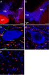
Figure 1.
Figure 1.
Comparative localization of α7 nAChRs (green), glutamatergic axon terminals (A-D, red; E, VGluT1, red, VGluT2, blue), and dopaminergic neurons (dendrites and cell bodies, blue) in aldehyde-fixed sections through the adult rat VTA. A-D, α7 nAChRs were labeled with Alexa Fluor 488-conjugated αBgt; glutamatergic axon terminals were labeled with anti-VGluT1 or -VGluT2, followed by Alexa Fluor 546-conjugated secondary antibody; dopaminergic neurons were labeled with anti-tyrosine hydroxylase, followed by Alexa Fluor 633-conjugated secondary antibody. E, VGluT1 and VGluT2 terminals were labeled with rabbit anti-VGluT1 and guinea pig anti-VGluT2, respectively, followed by Alexa Fluor 546-conjugated anti-rabbit IgG and Alexa Fluor 633-conjugated anti-guinea pig IgG. A, B, Low-magnification images of VGluT1 (A) and VGluT2 (B) immunoreactivites in the VTA and surrounding nuclei. VGluT1 labeling is particularly strong in the interpeduncular nucleus (ipn), with moderate levels in the VTA (A). In contrast, VGluT2 immunoreactivity is stronger in the VTA but apparently absent from the ipn (B). Note that no αBgt signal was detected at this magnification. C, Higher-magnification image of the VTA labeled for both VGluT1 and VGluT2. Three populations of αBgt binding sites are revealed, associated with VGluT-immunoreactive axon terminals (yellow arrows), dopaminergic neurons (blue arrows), and nondopaminergic cells (green arrows). Note that not all glutamatergic axon terminals express detectable levels of α7 nAChRs (red arrows). D, Competition control, in which preincubation with an excess of unconjugated αBgt obliterated fluorescent αBgt labeling. Note the lack of green and yellow fluorescent spots. E, Dual labeling for VGluT1 (rabbit anti-VGluT1, red) and VGluT2 (guinea pig anti-VGluT2, blue) immunoreactivity in the VTA; no detectable colocalization of the two VGluT isoforms within the same structures was observed. snc, Substantia nigra pars compacta. Scale bars: A, B, 200 μm; C-E, 20 μm.
Figure 2.
Figure 2.
Colocalization of α7 nAChRs and glutamatergic axon terminals in aldehyde-fixed sections through the adult rat VTA. α7 nAChRs were labeled with Alexa Fluor 488-conjugated αBgt (A, F, green); glutamatergic axon terminals were labeled with anti-VGluT1 (B) or anti-VGluT2 (G), followed by Alexa Fluor 546-conjugated secondary antibody (red); dopaminergic neurons were labeled with anti-tyrosine hydroxylase, followed by Alexa Fluor 633-conjugated secondary antibody (C, H, blue). D, I, Merged images. αBgt binding sites colocalized with both VGluT1-immunopositive (A-D) and VGluT2-immunopositive (F-I) axon terminals. Fluorescence profile analyses, along the paths indicated by white arrows in D and I, confirm the colocalization of αBgt with both VGluT1-immunoreactive (E) and VGluT2-immunoreactive (J) axonal boutons (see Materials and Methods). FI, Fluorescence intensity. Scale bars, 5 μm.
Figure 3.
Figure 3.
Localization of VGluT immunoreactivity, but not α7 nAChRs, within GFAP-positive astrocytes in aldehyde-fixed sections through the adult rat VTA. α7 nAChRs were labeled with Alexa Fluor 488-conjugated αBgt (green); glutamatergic axon terminals were labeled with anti-VGluT1 and -VGluT2, followed by Alexa Fluor 546-conjugated secondary antibody (red); astrocytes were labeled with anti-GFAP, followed by Alexa Fluor 633-conjugated secondary antibody (blue). A, VGluT immunoreactivity was observed within GFAP-positive structures, exemplified in B by the fluorescence profile analysis of line i. Note the absence of associated αBgt label. αBgt binding sites also colocalized with glutamatergic axon terminals, exemplified in C by the fluorescence profile analysis of line ii. FI, Fluorescence intensity. Scale bars, 10 μm.
Figure 4.
Figure 4.
Triple labeling of α7 nAChRs (green), cholinergic axon terminals (red), and dopaminergic dendrites (A) or axonal boutons (B, blue) in aldehyde-fixed sections through the adult rat VTA (A) and NAcc (B). α7 nAChRs were labeled with Alexa Fluor 488-conjugated αBgt; cholinergic axon terminals were labeled with anti-VAChT, followed by Alexa Fluor 546-conjugated secondary antibody; dopaminergic neurons were labeled with anti-tyrosine hydroxylase, followed by Alexa Fluor 633-conjugated secondary antibody. In the VTA, α7 nAChRs (green arrows) are located at sites distant from cholinergic axon terminals (red arrows). In contrast, in the NAcc, a subpopulation of αBgt binding sites are associated with VAChT immunoreactivity (yellow arrows). Scale bars, 10 μm.
Figure 5.
Figure 5.
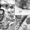
Synaptic localization of α7 nAChRs (arrows) in the adult rat VTA, revealed by labeling of unfixed slices. A, Unfixed slice incubated with biotinylated αBgt, followed by postembedding gold labeling for biotin. B, Magnified view of the boxed area in A, demonstrating the presence of gold particles (arrows) on the plasma membrane of an axonal bouton. C, Unfixed slice incubated directly with gold-conjugated αBgt. D, Magnified view of the boxed area in C, demonstrating the presence of gold particles (arrows) at a perisynaptic locus within a synaptic cleft. The silver reagent has fused the gold particles into a single unit, and, because this is in contact with both the presynaptic and postsynaptic membranes, it is not possible to determine on which membrane the α7 nAChRs are located. Arrows indicate αBgt binding sites. b, Axonal bouton; d, dendrite; pre, presynaptic bouton; post, postsynaptic dendrite. Scale bars: A-E, 100 nm; F, 150 nm.
Figure 6.
Figure 6.
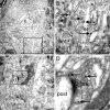
Subcellular localization of α7 nAChRs (some indicated by arrows) in the adult rat VTA, revealed by labeling of aldehyde-fixed sections. A, B, Sections incubated with biotinylated αBgt, followed by preembedding gold labeling for biotin. A, α7 nAChRs within a cell body in the VTA. B, Higher magnification of the boxed region in A reveals that a proportion of the αBgt binding sites are associated with the endoplasmic reticulum. C, D, Sections incubated directly with gold-conjugated αBgt. C, α7 nAChRs within an axonal bouton in the VTA. D, Higher magnification of the boxed region in C reveals that αBgt binding sites are located both within the cytoplasm and on the plasma membrane. Arrows indicate αBgt binding sites. b, Axonal bouton; c, cytoplasm; d, dendrite; er, endoplasmic reticulum; n, nucleus; pre, presynaptic bouton; post, postsynaptic dendrite. Scale bars: A, C, 500 nm; B, D, 100 nm.
Figure 7.
Figure 7.
Subcellular localization of α7 nAChRs (some indicated by arrows) in glutamatergic axon terminals (identified by VGluT immunoreactivity and revealed by the DAB reaction product) within aldehyde-fixed sections through the adult rat VTA. A, B, α7 nAChRs visualized with biotinylated αBgt in VGluT1-immunpositive (A) and VGluT2-immunopositive (B) axon terminals. C, D, α7 nAChRs visualized with gold-conjugated αBgt in VGluT1-immunopositive (C) and VGluT2-immunopositive (D) axon terminals. Arrows indicate αBgt binding sites. b, VGluT-immunoreactive axonal bouton; d, dendrite. Scale bars, 100 nm.
Figure 8.
Figure 8.
Subcellular relationship between α7 nAChRs and dopaminergic neurons (identified by tyrosine hydroxylase immunoreactivity and revealed by the DAB reaction product; A-C) or cholinergic axon terminals (identified by VAChT immunoreactivity and revealed by the DAB reaction product; D) within aldehyde-fixed sections through the adult rat VTA. A, α7 nAChRs, visualized with gold-conjugated αBgt, within the cytoplasm of a dopaminergic neuron. B, High-contrast image of the boxed area in A, facilitating visualization of the gold particles. C, Postsynaptic α7 nAChRs, visualized with gold-conjugated αBgt, on the plasma membrane of a dopaminergic neuron. D, VAChT-immunoreactive axon terminal within the VTA. Note that, although gold-conjugated αBgt labeling is present (arrow), it is not associated with the VAChT-positive terminal. Arrows indicate αBgt binding sites. b, Axonal bouton; c, cytoplasm; d, dendrite. Scale bars: A, 500 nm; B, D, 200 nm; C, 100 nm.

References

    1. Alkondon M, Rocha ES, Maelicke A, Albuquerque EX (1996) Diversity of nicotinic acetylcholine receptors in rat brain. V. α-Bungarotoxin-sensitive nicotinic receptors in olfactory bulb neurons and presynaptic modulation of glutamate release. J Pharmacol Exp Ther 278: 1460-1471. - PubMed
    1. Alkondon M, Pereira EF, Cortes WS, Maelicke A, Albuquerque EX (1997) Choline is a selective agonist of α7 nicotinic acetylcholine receptors in the rat brain neurons. Eur J Neurosci 9: 2734-2742. - PubMed
    1. Aramakis VB, Metherate R (1998) Nicotine selectively enhances NMDA receptor-mediated synaptic transmission during postnatal development in sensory neocortex. J Neurosci 18: 8485-8495. - PMC - PubMed
    1. Bezzi P, Gundersen V, Galbete JL, Seifert G, Steinhauser C, Pilati E, Volterra A (2004) Astrocytes contain a vesicular compartment that is competent for regulated exocytosis of glutamate. Nat Neurosci 7: 613-620. - PubMed
    1. Caruncho HJ, Guidotti A, Lindstrom J, Costa E, Pesold C (1997) Subcellular localization of the α7 nicotinic receptor in rat cerebellar granule cell layer. NeuroReport 8: 1431-1433. - PubMed

Publication types

MeSH terms

LinkOut - more resources