. 2004 Dec 15;24(50):11337-45.
doi: 10.1523/JNEUROSCI.3698-04.2004.

Conversion of the modulatory actions of dopamine on spinal reflexes from depression to facilitation in D3 receptor knock-out mice

Affiliations

Conversion of the modulatory actions of dopamine on spinal reflexes from depression to facilitation in D3 receptor knock-out mice

Stefan Clemens et al. J Neurosci. .

Abstract

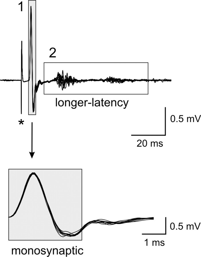
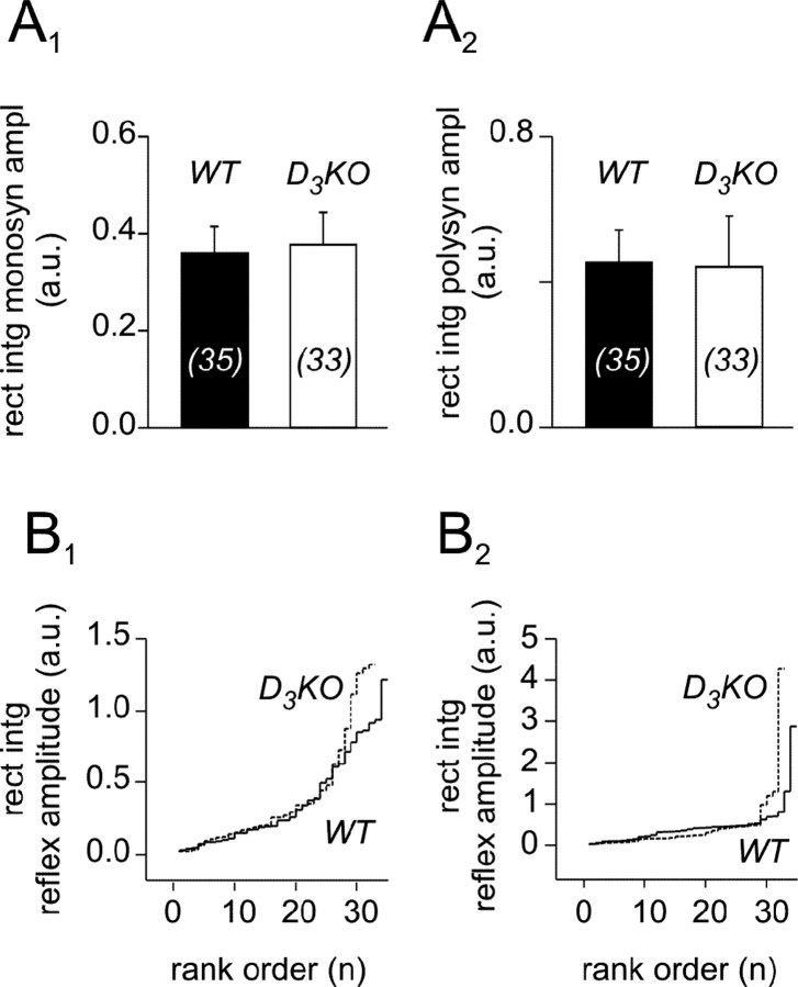
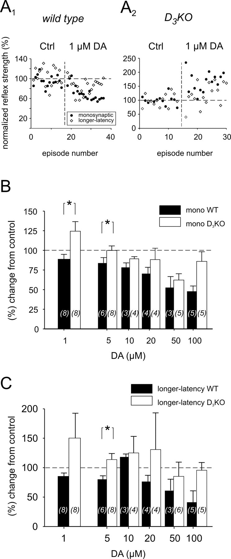
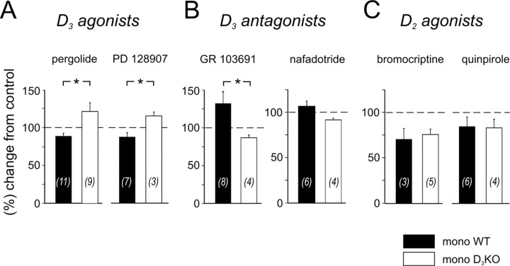
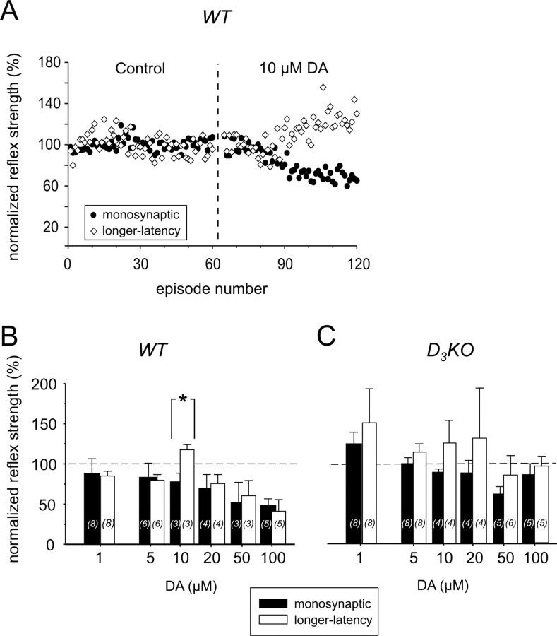
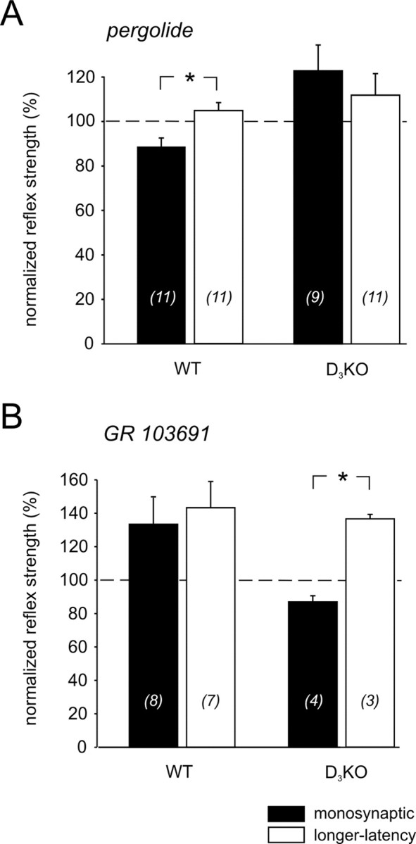
Descending monoaminergic systems modulate spinal cord function, yet spinal dopaminergic actions are poorly understood. Using the in vitro lumbar cord, we studied the effects of dopamine and D2-like receptor ligands on spinal reflexes in wild-type (WT) and D3-receptor knock-out mice (D3KO). Low dopamine levels (1 microM) decreased the monosynaptic "stretch" reflex (MSR) amplitude in WT animals and increased it in D3KO animals. Higher dopamine concentrations (10-100 microM) decreased MSR amplitudes in both groups, but always more strongly in WT. Like low dopamine, the D3 receptor agonists pergolide and PD 128907 reduced MSR amplitude in WT but not D3KO mice. Conversely, D3 receptor antagonists (GR 103691 and nafadotride) increased the MSR in WT but not in D3KO mice. In comparison, D2-preferring agonists bromocriptine and quinpirole depressed the MSR in both groups. Low dopamine (1-5 microM) also depressed longer-latency (presumably polysynaptic) reflexes in WT but facilitated responses in D3KO mice. Additionally, in some experiments (e.g., during 10 microM dopamine or pergolide in WT), polysynaptic reflexes were facilitated in parallel to MSR depression, demonstrating differential modulatory control of these reflex circuits. Thus, low dopamine activates D3 receptors to limit reflex excitability. Moreover, in D3 ligand-insensitive mice, excitatory actions are unmasked, functionally converting the modulatory action of dopamine from depression to facilitation. Restless legs syndrome (RLS) is a CNS disorder involving abnormal limb sensations. Because RLS symptoms peak at night when dopamine levels are lowest, are relieved by D3 agonists, and likely involve increased reflex excitability, the D3KO mouse putatively explains how impaired D3 activity could contribute to this sleep disorder.


Figures

Figure 1.
Figure 1.
Example of a reflex response recorded from the ventral roots (10 consecutive sweeps superimposed). Stimulation of an L5 dorsal root (500 μA, 500 μsec, at asterisk) induced a reflex response in the corresponding ventral root that consisted of a monosynaptic (1) and a longer-latency reflex response (2). Note that the longer-latency response consists here of two epochs that can be easily distinguished, at ∼15-30 and ∼40-60 msec after the onset of the MSR, respectively. The boxed regions identify those epochs chosen to calculate the amplitude of monosynaptic and longer-latency reflexes. The MSR component is expanded horizontally to highlight the 3 msec period and show its stability.
Figure 2.
Figure 2.
The MSR and longer-latency reflex amplitudes of WT and D3KO mice have a similar average but a slightly different distribution pattern. A, Comparison of the rectified and integrated amplitudes. A1, Monosynaptic reflex amplitudes of wild-type and D3KO animals under controlconditions. In WT animals, the average is 0.36 ± 0.05 mV (n = 35), and in D3KO animals it is 0.38 ± 0.07 mV (n = 33). The populations are not different (p = 0.932). A2, Longer-latency reflex amplitudes of WT and D3KO animals under control conditions. In WT, the average is 0.44 ± 0.08 mV, and in D3KO animals it is 0.43 ± 0.13 mV. Again, there is no significant difference between the two populations (p = 0.948). Sample sizes are indicated within histogram bars in brackets. B, Rank ordered histograms of WT and D3KO reflex responses. Data are from the same pool as in A and plotted as a function of their rank. B1, Monosynaptic reflex amplitudes. WT (solid line) and D3KO (dashed line) reflex amplitudes are similar for low and median ranks, but there are more larger-reflex amplitudes in D3KO than in WT. B2, Longer-latency reflex amplitudes. Here again, WT (solid line) and D3KO (dashed line) reflex amplitudes are similar for low and median ranks, and there are more larger-reflex amplitudes in D3KO than in WT animals. rect in t monosyn ampl, Rectified and integrated monosynaptic reflex amplitude; polysyn ampl, rectified and integrated polysynaptic reflex amplitude; a.u., artificial units.
Figure 3.
Figure 3.
Effects of dopamine (DA) on MSR (mono) and longer-latency reflex amplitudes in WT and D3KO animals. A1, Typical example of a WT animal, in which application of 1 μm dopamine to the bath led to a depression of monosynaptic and longer-latency reflex amplitudes. A2, Example of the effect of bath application of 1 μm dopamine to a D3KO animal preparation. Here, MSR and longer-latency amplitude increased during dopamine application. B, Comparison of the dopamine effects on the MSR amplitude at increasing concentrations. At 1 μm dopamine, the MSR amplitude of WT animals decreased, whereas it increased in D3KO animals. The difference between WT and D3KO mice was significant (p = 0.021). At 5 μm dopamine, there was still a significant difference between the two groups (p = 0.013); however, the average amplitude in the D3KO animals was no longer facilitated over the control response. At 10-50 μm dopamine, the reflex amplitudes of both animal types were depressed similarly, although the WT continued to show a slightly stronger depression than the D3KO animals. At 100 μm dopamine, however, D3KO animals showed a noticeably smaller depression than the WT animals. C, Comparison of the dopamine effects on longer-latency reflex amplitudes at increasing dopamine concentrations. At 1 and 5 μm dopamine, the reflex amplitude of WT animals generally decreased, whereas the amplitude of D3KO animals increased. This difference between WT and D3KO mice was significant at 5 μm (p = 0.026). At 10-100 μm dopamine, the depression observed in WT animals was consistently slightly stronger than in the D3KO animals. In this and the following figures, sample sizes are indicated within histogram bars in brackets. All comparisons are based on ANOVA with Tukey or Dunn's post hoc comparison. Asterisks denote significant differences.
Figure 4.
Figure 4.
Differential actions of D3 receptor ligands on the MSR amplitudes in WT and D3KO animals. A, D3 receptor agonists. Pergolide (left) induced a depression of the amplitude in WT animals (to 89 ± 4%) but led to a facilitation of the reflex amplitude in the D3KO animals (to 121 ± 12%), which was significantly different (p = 0.012). Similarly, PD 128907 (right) also induced a depression of the monosynaptic amplitude in the WT animals (to 88 ± 6%) and a facilitation in the D3KO animals (to 123 ± 16%). Here again, the differences between the modulatory effects was significant (p = 0.033). B, D3 receptor antagonists. GR 103691 (left) induced a facilitation of the amplitude in WT animals (to 134 ± 15% of control) but led to a depression of the reflex amplitude in the D3KO animals (to 87 ± 4% of control). This difference was significant (p = 0.029). Similarly, nafadotride (right) also induced a facilitation of the monosynaptic amplitude in the WT animals (to 107 ± 6%), and a depression in the D3KO animals (to 92 ± 2%); however, the overall difference was not significant (p = 0.067). C, D2 receptor agonists. Bromocriptine (left) induced a depression of the amplitude in WT animals (to 70.1 ± 11.8% of control) and D3KO animals (to 75.4 ± 5.9% of control) alike (p = 1.0). Similarly, quinpirole (right) also induced a depression of the monosynaptic amplitude in both WT animals (to 84.3 ± 10.4%) and D3KO animals (to 82.8 ± 9.5%), which again was not different (p = 0.914).
Figure 5.
Figure 5.
Monosynaptic and longer-latency reflex responses can be modulated differently by dopamine. A, Example of a differential modulation of monosynaptic and longer-latency reflex responses in the presence of 10 μm dopamine. Dopamine induced a depression of the MSR, yet at the same time also evoked a facilitation of the longer-latency reflex. B, In WT animals, the differential modulation of monosynaptic and longer-latency reflex amplitude was significant only at a concentration of 10 μm dopamine (p = 0.01; ANOVA with Tukey post hoc comparison), but not at the lower and higher concentrations tested. C, In contrast, in D3KO animals, we did not observe a similar differential modulation of monosynaptic and longer-latency reflexes within the range of dopamine concentrations tested. Note, however, the comparatively greater variability of reflex amplitudes in the D3KO animals. DA, Dopamine.
Figure 6.
Figure 6.
Differential actions of D3 receptor-specific drugs on the monosynaptic and longer-latency reflex amplitudes in WT and D3KO animals. A, The D3 receptor agonist pergolide induced a depression of the monosynaptic amplitude (black) in WT animals (to 89 ± 4% of control), but also led to a facilitation of the longer-latency reflex amplitude (white) (to 105 ± 4% of control; p = 0.007). There was no such difference in the effects of pergolide in D3KO animals. B, Conversely, in D3KO but not WT animals, the D3 receptor antagonist GR 103691 induced a depression of the monosynaptic amplitude (to 87 ± 4%) and a facilitation of the longer-latency reflex amplitude (to 137 ± 3%), which also was different (p < 0.001).

References

    1. Abramets II, Samoilovich IM (1991) Analysis of two types of dopaminergic responses of neurons of the spinal ganglia of rats. Neurosci Behav Physiol 21: 435-440. - PubMed
    1. Accili D, Fishburn CS, Drago J, Steiner H, Lachowicz JE, Park BH, Gauda EB, Lee EJ, Cool MH, Sibley DR, Gerfen CR, Westphal H, Fuchs S (1996) A targeted mutation of the D3 dopamine receptor gene is associated with hyperactivity in mice. Proc Natl Acad Sci USA 93: 1945-1949. - PMC - PubMed
    1. Ali NJ, Davies RJ, Fleetham JA, Stradling JR (1991) Periodic movements of the legs during sleep associated with rises in systemic blood pressure. Sleep 14: 163-165. - PubMed
    1. Allen RP, Earley CJ (2001) Restless legs syndrome: a review of clinical and pathophysiologic features. J Clin Neurophysiol 18: 128-147. - PubMed
    1. Asico LD, Ladines C, Fuchs S, Accili D, Carey RM, Semeraro C, Pocchiari F, Felder RA, Eisner GM, Jose PA (1998) Disruption of the dopamine D3 receptor gene produces renin-dependent hypertension. J Clin Invest 102: 493-498. - PMC - PubMed

Publication types

MeSH terms