

Localization of brain-derived neurotrophic factor to distinct terminals of mossy fiber axons implies regulation of both excitation and feedforward inhibition of CA3 pyramidal cells
- PMID: 15601941
- PMCID: PMC1351361
- DOI: 10.1523/JNEUROSCI.3846-04.2004
Localization of brain-derived neurotrophic factor to distinct terminals of mossy fiber axons implies regulation of both excitation and feedforward inhibition of CA3 pyramidal cells
Abstract
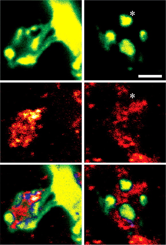
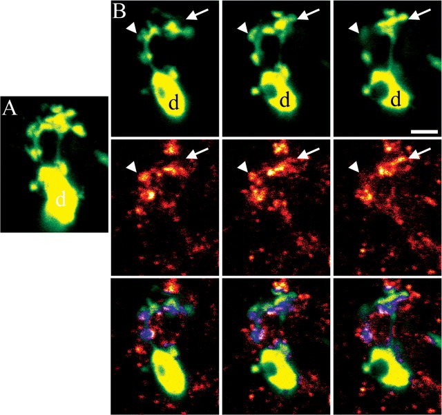
Hippocampal dentate granule cells directly excite and indirectly inhibit CA3 pyramidal cells via distinct presynaptic terminal specializations of their mossy fiber axons. This mossy fiber pathway contains the highest concentration of brain-derived neurotrophic factor (BDNF) in the CNS, yet whether BDNF is positioned to regulate the excitatory and/or inhibitory pathways is unknown. To localize BDNF, confocal microscopy of green fluorescent protein transgenic mice was combined with BDNF immunohistochemistry. Approximately half of presynaptic granule cell-CA3 pyramidal cell contacts were found to contain BDNF. Moreover, enhanced neuronal activity virtually doubled the percentage of BDNF-immunoreactive terminals contacting CA3 pyramidal cells. To our surprise, BDNF was also found in mossy fiber terminals contacting inhibitory neurons. These studies demonstrate that mossy fiber BDNF is poised to regulate both direct excitatory and indirect feedforward inhibitory inputs to CA3 pyramdal cells and reveal that seizure activity increases the pool of BDNF-expressing granule cell presynaptic terminals contacting CA3 pyramidal cells.
Figures


Similar articles
-
Altered morphology of hippocampal dentate granule cell presynaptic and postsynaptic terminals following conditional deletion of TrkB.Hippocampus. 2008;18(7):668-78. doi: 10.1002/hipo.20426. Hippocampus. 2008. PMID: 18398849 Free PMC article.
-
Structural plasticity of dentate granule cell mossy fibers during the development of limbic epilepsy.Hippocampus. 2010 Jan;20(1):113-24. doi: 10.1002/hipo.20589. Hippocampus. 2010. PMID: 19294647 Free PMC article.
-
Prenatal hippocampal granule cells in primary cell culture form mossy fiber boutons at pyramidal cell dendrites.J Neurosci Res. 1998 Mar 1;51(5):602-11. doi: 10.1002/(SICI)1097-4547(19980301)51:5<602::AID-JNR7>3.0.CO;2-J. J Neurosci Res. 1998. PMID: 9512004
-
Interneuron diversity series: containing the detonation--feedforward inhibition in the CA3 hippocampus.Trends Neurosci. 2003 Nov;26(11):631-40. doi: 10.1016/j.tins.2003.09.007. Trends Neurosci. 2003. PMID: 14585604 Review.
-
Synapses formed by normal and abnormal hippocampal mossy fibers.Cell Tissue Res. 2006 Nov;326(2):361-7. doi: 10.1007/s00441-006-0269-2. Epub 2006 Jul 4. Cell Tissue Res. 2006. PMID: 16819624 Review.
Cited by
-
BDNF and its pro-peptide are stored in presynaptic dense core vesicles in brain neurons.J Cell Biol. 2012 Mar 19;196(6):775-88. doi: 10.1083/jcb.201201038. Epub 2012 Mar 12. J Cell Biol. 2012. PMID: 22412021 Free PMC article.
-
Visualizing BDNF Transcript Usage During Sound-Induced Memory Linked Plasticity.Front Mol Neurosci. 2018 Jul 31;11:260. doi: 10.3389/fnmol.2018.00260. eCollection 2018. Front Mol Neurosci. 2018. PMID: 30127717 Free PMC article.
-
TrkB-mediated activation of the phosphatidylinositol-3-kinase/Akt cascade reduces the damage inflicted by oxygen-glucose deprivation in area CA3 of the rat hippocampus.Eur J Neurosci. 2018 May;47(9):1096-1109. doi: 10.1111/ejn.13880. Epub 2018 Mar 25. Eur J Neurosci. 2018. PMID: 29480936 Free PMC article.
-
Differential regulation of BDNF, synaptic plasticity and sprouting in the hippocampal mossy fiber pathway of male and female rats.Neuropharmacology. 2014 Jan;76 Pt C(0 0):696-708. doi: 10.1016/j.neuropharm.2013.04.029. Epub 2013 May 6. Neuropharmacology. 2014. PMID: 23660230 Free PMC article.
-
Activity-dependent BDNF release and TRPC signaling is impaired in hippocampal neurons of Mecp2 mutant mice.Proc Natl Acad Sci U S A. 2012 Oct 16;109(42):17087-92. doi: 10.1073/pnas.1205271109. Epub 2012 Oct 1. Proc Natl Acad Sci U S A. 2012. PMID: 23027959 Free PMC article.
References
-
- Alderson RF, Curtis R, Alterman AL, Lindsay RM, DiStefano PS (2000) Truncated TrkB mediates the endocytosis and release of BDNF and neurotrophin-4/5 by rat astrocytes and Schwann cells in vitro. Brain Res 871: 210-222. - PubMed
-
- Altar CA, Siuciak JA, Wright P, Ip NY, Lindsay RM, Wiegand SJ (1994) In situ hybridization of trkB and trkC receptor mRNA in rat forebrain and association with high-affinity binding of [125I]BDNF, [125I]NT-4/5 and [125I]NT-3. Eur J Neurosci 6: 1389-1405. - PubMed
-
- Amaral DG (1979) Synaptic extensions from the mossy fibers of the fascia dentata. Anat Embryol (Berl) 155: 241-251. - PubMed
-
- Amaral DG, Dent JA (1981) Development of the mossy fibers of the dentate gyrus: I. A light and electron microscopic study of the mossy fibers and their expansions. J Comp Neurol 195: 51-86. - PubMed
Publication types
MeSH terms
Substances
Grants and funding
LinkOut - more resources
Full Text Sources
Other Literature Sources
Miscellaneous