. 2004 Dec 15;24(50):11439-48.
doi: 10.1523/JNEUROSCI.3459-04.2004.

Interaction between the corticotropin-releasing factor system and hypocretins (orexins): a novel circuit mediating stress response

Affiliations

Interaction between the corticotropin-releasing factor system and hypocretins (orexins): a novel circuit mediating stress response

Raphaëlle Winsky-Sommerer et al. J Neurosci. .

Abstract

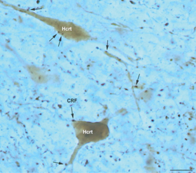
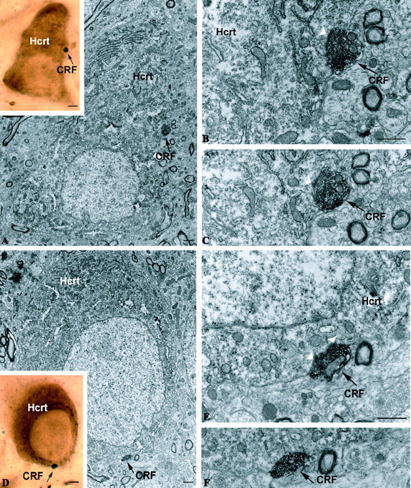
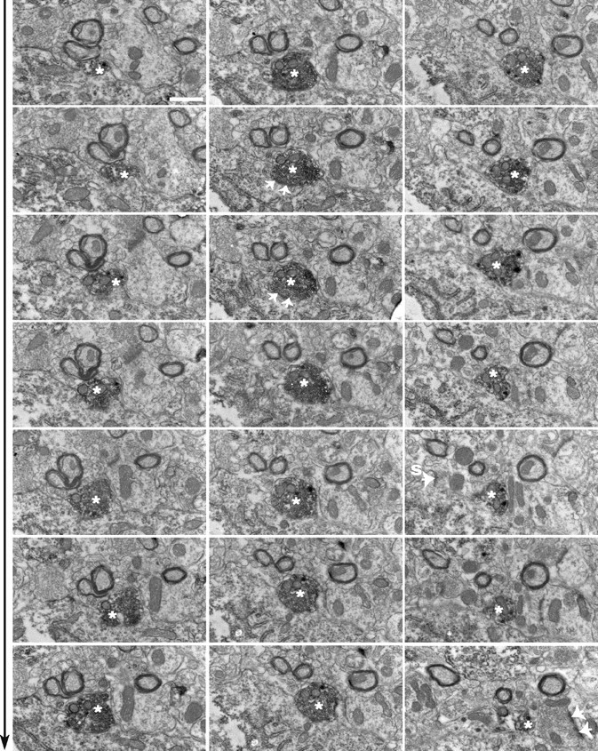
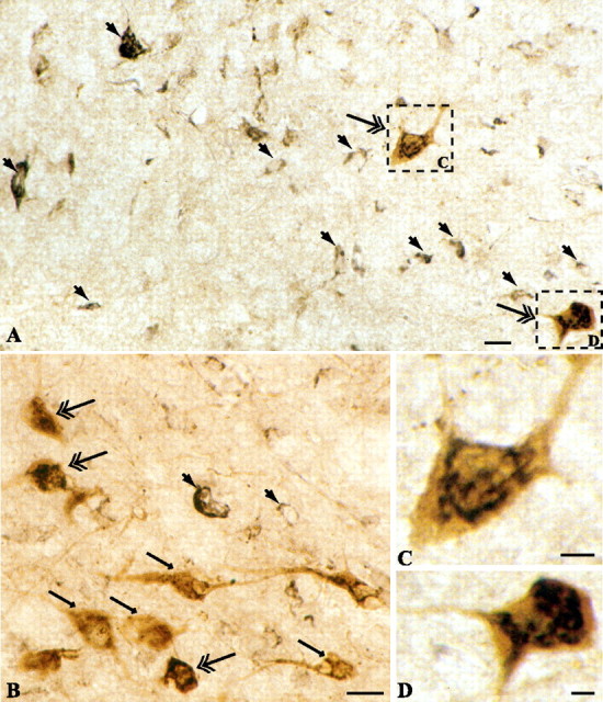
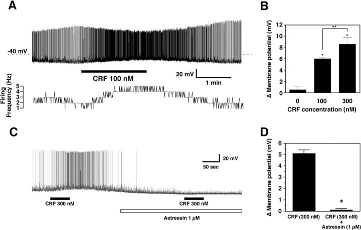
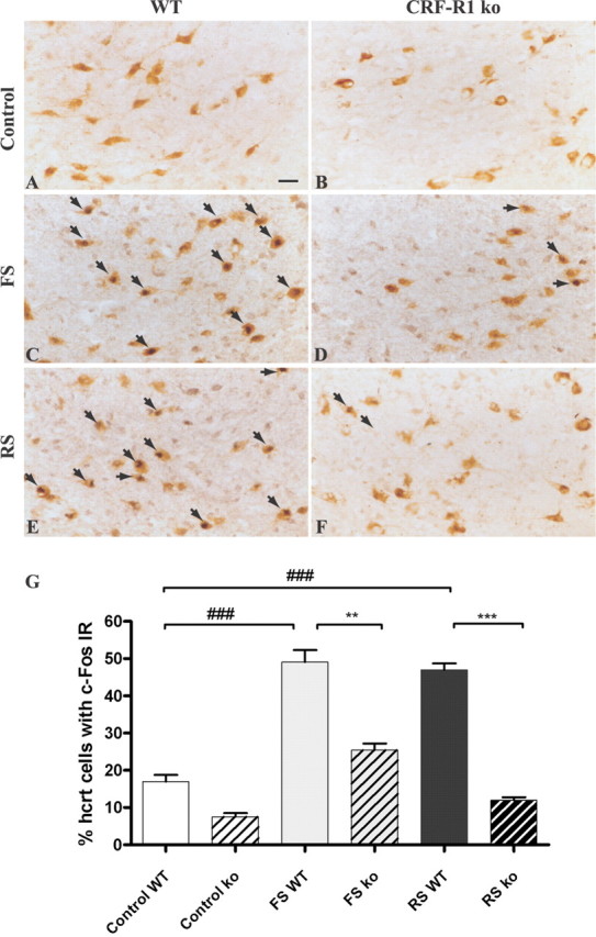
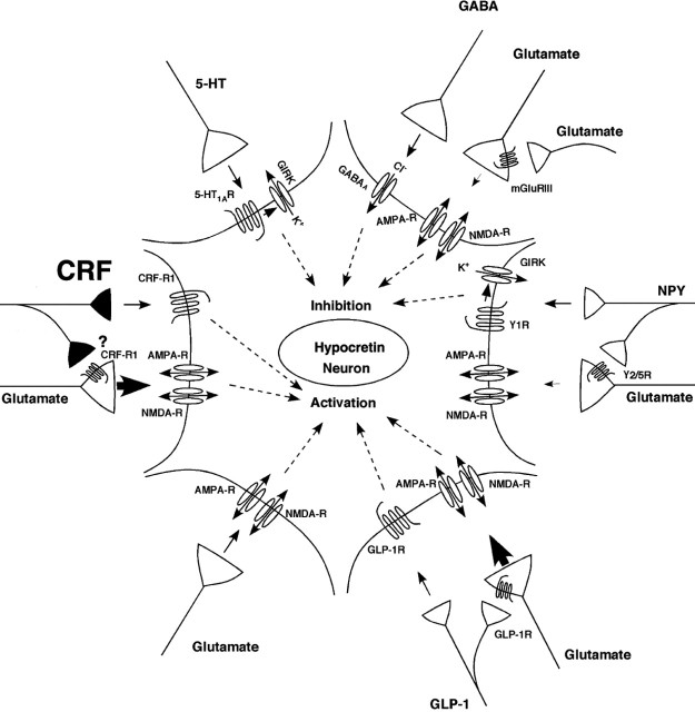
The hypothalamic neuropeptides hypocretins (orexins) play a crucial role in the stability of arousal and alertness. We tested whether the hypocretinergic system is a critical component of the stress response activated by the corticotropin-releasing factor (CRF). Our results show that CRF-immunoreactive terminals make direct contact with hypocretin-expressing neurons in the lateral hypothalamus and that numerous hypocretinergic neurons express the CRF-R1/2 receptors. We also demonstrate that application of CRF to hypothalamic slices containing identified hypocretin neurons depolarizes membrane potential and increases firing rate in a subpopulation of hypocretinergic cells. CRF-induced depolarization was tetrodotoxin insensitive and was blocked by the peptidergic CRF-R1 antagonist astressin. Moreover, activation of hypocretinergic neurons in response to acute stress was severely impaired in CRF-R1 knock-out mice. Together, our data provide evidence of a direct neuroanatomical and physiological input from CRF peptidergic system onto hypocretin neurons. We propose that, after stressor stimuli, CRF stimulates the release of hypocretins and that this circuit contributes to activation and maintenance of arousal associated with the stress response.


Figures

Figure 1.
Figure 1.
Putative contacts between CRF bouton-like structures and hypocretinergic neurons in the lateral hypothalamus. CRF-immunoreactive boutons (arrow; DAB-nickel) were observed in close apposition to hypocretin-expressing perikarya and dendrites (DAB). Scale bar, 10 μm.
Figure 2.
Figure 2.
Synaptic interaction between CRF axons and hypocretin. A-C, D-F, Correlated light and electron micrographs demonstrating black-labeled CRF axon terminals (black arrow) synapsing on hypocretin-immunoreactive perikarya. A and D are the light micrographs of the putative contacts. Scale bar (in A, D), 2 μm. B, C, E, F, Electron micrograph showing an asymmetrical synaptic membrane specialization (white arrowheads) between the same CRF bouton-like structure (A, D) and the hypocretin-immunolabeled perikaryon in the lateral hypothalamus. Scale bars: B, E, 1 μm.
Figure 3.
Figure 3.
Serial sections of a CRF axon terminal on hcrt perikaryon. Electron micrographic panels (from left to right, top to bottom in sequence) showing the emergence and dissipation of a CRF-immunoreactive bouton (white asterisk) in association with an hcrt perikaryon. This same interaction was depicted on correlated light and electron micrographs of Figure 2 D-F. Note that the orientation of the panels in this figure is 90° counterclockwise rotated compared with their position on Figure 2. The CRF-immunoreactive bouton establishes an asymmetrical contact with the hcrt perikarya (white arrows). The same bouton is also in direct contact with another presynaptic bouton, which itself establishes an asymmetrical contact on a dendritic shaft. Large-cored vesicles immunolabeled for CRF appear in the vicinity of this unlabeled presynaptic terminal. This ultrastructural substrate is consistent with the observed effect of TTX on CRF-induced depolarization of hcrt neurons, suggesting both direct postsynaptic and presynaptic effects of CRF. Another interesting feature of hypocretinergic perikarya was the presence of somatic spines, which received asymmetrical contacts (white arrow). Scale bar, 1 μm.
Figure 4.
Figure 4.
Specificity controls of CRF-R1/2 immunostaining in the brain. Sections at the level of the hippocampus (A) and paraventricular nucleus of the hypothalamus (C) display positive immunofluorescent staining with CRF-R1/2 antibody. Immunoreactive signals are no longer apparent when the antiserum was preadsorbed with the immunogenic peptide (B, D). Scale bars: A, B, 200 μm; C, D, 80 μm; (in E) E-G, 10 μm. E-G, Double labeling of tissue sections at the level of the lateral hypothalamus, respectively, showing the CRF-R1/2 (E) and the hypocretin (F) immunoreactivities. F, Merged image of E and F, illustrating the colocalization (yellow signal, arrow) between CRF-R1/2 (green) and hypocretin (red).
Figure 5.
Figure 5.
Hypocretin-producing cells express CRF receptors. A, B, Neurons in the perifornical region of the lateral hypothalamus express CRF receptors (large solid arrow; black reaction product). Double labeling shows that numerous hypocretin-immunopositive perikarya displayed CRF-R1/2 immunoreactivity (double arrow). Single-labeled hypocretin-expressing neurons are shown by arrow (brown reaction product). Scale bars: A, B, 10 μm. C, D, Higher magnification of two hypocretinergic neurons (frames in A) illustrating colocalization of hypocretin and CRF-R1 immunoreactivities. Scale bars: C, D, 2.5 μm.
Figure 6.
Figure 6.
Response of orexin/EGFP neurons to CRF application. A, Depolarization and increased firing of an orexin/EGFP neuron result from 100 nm application of CRF. Membrane potential and firing rate return to basal levels several minutes after washout begins. B, Change in membrane potential is dependent on CRF concentration. Values are mean ± SEM; * p < 0.002 versus control; **p < 0.05 for 100 versus 300 nm. C, The depolarizing effect of CRF is blocked in the presence of the CRF-R1 antagonist astressin. D, The CRF-induced change in membrane potential is significantly reduced (*p = 0.0003) in the presence of the CRF-R1 antagonist. Values are mean ± SEM.
Figure 7.
Figure 7.
Activation of hypocretin neurons during footshock and restraint stress in CRF receptor 1 knock-out versus control animals. A-F, Representative tissue sections at the level of the perifornical area of the dorsolateral hypothalamus illustrating hypocretin (brown DAB staining) and c-Fos (dark blue DAB-NiCl2) immunoreactivities for control groups (A, B) and stressed groups (C-F). Double-labeled neurons are shown by arrows. Scale bar (in A), A-F, 10 μm. E, Percentage of hypocretinergic neurons immunoreactive for c-Fos in CRF-R1 knock-out (ko) versus wild-type (WT) mice during footshock (FS) or restraint stress (RS) challenge. Data are expressed as mean ± SEM. **p < 0.001; ***,# # #p < 0.0001. n.s., Not significant.
Figure 8.
Figure 8.
Schematic illustrating electrophysiologically demonstrated inputs to hypocretin neurons. The present study demonstrates that CRF can depolarize a subset of these neurons through the CRF-R1. Other inputs are not illustrated because the receptor mediating the response has yet to be determined, including noradrenergic and cholinergic inputs and other factors such as leptin, glucose, and ghrelin.

References

    1. Acuna-Goycolea C, van den Pol A (2004) Glucagon-like peptide 1 excites hypocretin/orexin neurons by direct and indirect mechanisms: implications for viscera-mediated arousal. J Neurosci 24: 8141-8152. - PMC - PubMed
    1. Acuna-Goycolea C, Li Y, van Den Pol AN (2004) Group III metabotropic glutamate receptors maintain tonic inhibition of excitatory synaptic input to hypocretin/orexin neurons. J Neurosci 24: 3013-3022. - PMC - PubMed
    1. Alam MN, Gong H, Alam T, Jaganath R, McGinty D, Szymusiak R (2002) Sleep-waking discharge patterns of neurons recorded in the rat perifornical lateral hypothalamic area. J Physiol (Lond) 538: 619-631. - PMC - PubMed
    1. Blanco M, Lopez M, GarcIa-Caballero T, Gallego R, Vazquez-Boquete A, Morel G, SenarIs R, Casanueva F, Dieguez C, Beiras A (2001) Cellular localization of orexin receptors in human pituitary. J Clin Endocrinol Metab 86: 1616-1619. - PubMed
    1. Blanco M, Gallego R, Garcia-Caballero T, Dieguez C, Beiras A (2003) Cellular localization of orexins in human anterior pituitary. Histochem Cell Biol 120: 259-264. - PubMed

Publication types

MeSH terms