. 2004 Dec 15;32(22):6490-500.
doi: 10.1093/nar/gkh987. Print 2004.

Rad23 stabilizes Rad4 from degradation by the Ub/proteasome pathway

Affiliations

Rad23 stabilizes Rad4 from degradation by the Ub/proteasome pathway

Tatiana G Ortolan et al. Nucleic Acids Res. .

Abstract

Rad23 protein interacts with the nucleotide excision-repair (NER) factor Rad4, and the dimer can bind damaged DNA. Rad23 also binds ubiquitinated proteins and promotes their degradation by the proteasome. Rad23/proteasome interaction is required for efficient NER, although the specific role of the Ub/proteasome system in DNA repair is unclear. We report that the availability of Rad4 contributes significantly to the cellular tolerance to UV light. Mutations in the proteasome, and in genes encoding the ubiquitin-conjugating enzymes Ubc4 and Ubc5, stabilized Rad4 and increased tolerance to UV light. A short amino acid sequence, previously identified in human Rad23, mediates the interaction between Rad23 and Rad4. We determined that this motif was required for stabilizing Rad4, and could function independently of the intact protein. A ubiquitin-like (UbL) domain in Rad23 binds the proteasome, and is required for conferring full resistance to DNA damage. However, Rad23/proteasome interaction appears unrelated to Rad23-mediated stabilization of Rad4. Specifically, simultaneous expression of a Rad23 mutant that could not bind the proteasome, with a mutant that could not interact with Rad4, fully suppressed the UV sensitivity of rad23Delta, demonstrating that Rad23 performs two independent, but concurrent roles in NER.


Figures
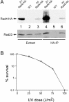
Figure 1
Figure 1
Physiological expression of an epitope-tagged derivative of Rad4. (A) The native RAD4 gene was replaced with a version containing two tandem HA epitopes. Protein extracts were prepared and equal amounts were resolved by SDS–PAGE. A nitrocellulose filter was incubated with antibodies against HA and Rad4–HA was detected when expressed from a high copy plasmid (lane 1) and at physiological levels from the integrated gene (lane 3). Protein extracts were also incubated with anti-HA antibodies to immunoprecipitate Rad4–HA, and verify interaction with endogenous Rad23. High levels of Rad4–HA were recovered from cells expressing the protein from a high-copy plasmid, in contrast to ∼25-fold lower levels from the integrated derivative. Rad23 was co-precipitated with Rad4–HA from both strains (lanes 4 and 6). Significantly, neither Rad4–HA nor Rad23 was purified from a strain that expressed untagged Rad4 (lane 5). (B) To confirm that Rad4–HA provided UV resistance, yeast cells were diluted to ∼1 × 10⁷/ml and spread evenly on agarose medium. The plates were exposed to 254 nm UV light for the doses indicated, covered with foil, and incubated at 30°C for ∼3 days. The number of colonies was counted from duplicate experiments, and the average values are plotted. (Wild-type, gray square; Rad4–HA integrant, solid diamond.)
Figure 2
Figure 2
Rad23 regulates Rad4–HA abundance. (A) Wild-type and rad23Δ cells expressing physiological levels of Rad4–HA were incubated with 35S-methionine + 35S-cysteine for 10 min. The cells were suspended in medium containing excess unlabeled amino acids and cycloheximide. Extracts were prepared at the intervals indicated and Rad4–HA was immunoprecipitated (arrow) and detected by autoradiography. The major band at ∼70 kDa is a non-specific interaction. The steady-state levels of Rad4–HA that was expressed from a high-copy plasmid (B) was compared to the expression from an integrated gene (C) in RAD23 and rad23Δ cells. An aliquot was withdrawn from the culture before translation inhibitor was added (Con). Following addition of cycloheximide, aliquots were withdrawn immediately (0), and at the intervals indicated. Total protein was examined by SDS–PAGE and immunoblotting with anti-HA antibodies. Equality of protein loading was confirmed by incubating the same blot with antibodies against Pab1.
Figure 3
Figure 3
Episomal Rad23 can restore Rad4 levels in rad23Δ cells. (A) A plasmid encoding FLAG–Rad23 was transformed into wild-type and rad23Δ cells that also contained integrated Rad4–HA. The levels of Rad4–HA was determined by immunoblotting. Rad4–HA levels were restored to wild-type levels following transformation of the Rad23-encoding plasmid (compare lanes 1 and 3). FLAG–Rad23 is slightly larger than the native protein, and this difference is observed in SDS–PAGE. To confirm equality of loading, the same filters were incubated with antibodies against eEF1A. (B) The stability of Rad4–HA, expressed at high levels from a PCUP1-driven promoter, was examined by pulse-labeling methods in cells containing or lacking Rad23. Aliquots of the labeled extracts were withdrawn at the times indicated, precipitated with anti-HA antibodies, and following SDS–PAGE, examined by autoradiography. The amount of Rad4–HA remaining over time is indicated at the bottom of the panel. (C) The levels of endogenous Rad23 were examined in yeast cells that expressed different levels of untagged Rad4. Three different loadings of total protein extract (5, 10 and 20 μg) were prepared from RAD4, rad4Δ and PCUP1::RAD4-over-expressing cells and resolved by SDS–PAGE. An immunoblot was incubated with antibodies against Rad23.
Figure 4
Figure 4
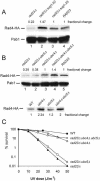
Rad4 levels are regulated by the Ub/proteasome system. (A) The levels of Rad4–HA were measured in the sug1 proteasome mutant strain, and compared to wild-type and rad23Δ cells. The values above the panel indicate relative amounts of Rad4–HA that was measured by densitometry. (B) The decreased levels of Rad4–HA in rad23Δ are fully restored in a yeast mutant lacking the Ub-conjugating enzymes Ubc4 and Ubc5 (quantitation above the panel). The lower panel shows that Rad4–HA levels are also increased in a RAD23 strain that lacks Ubc4 (or both Ubc4 and Ubc5; quantitation below the panel). (C) Yeast cells lacking Ubc4, or both Ubc4 and Ubc5, can strongly suppress the UV-sensitivity of rad23Δ. Although Ubc4 and Ubc5 are highly homologous, Ubc5 is expressed at very low levels, and primarily in growth-arrested cells. (Wild-type, solid square; rad23Δ, solid triangle; rad23Δ ubc5Δ, open square; rad23Δ ubc4Δ, open triangle; rad23Δ ubc4Δ ubc5Δ, open circle.)
Figure 5
Figure 5
The Rad4-binding (R4B) domain in Rad23 is required for stabilizing Rad4–HA. (A) A scheme that indicates the various domains of Rad23 is shown. FLAG-tagged derivatives lacking the N-terminal UbL sequence, or the R4B sequence are shown. A derivative containing two single amino acid substitutions, in the two UBA domains, is also shown. (B) The DNA repair proficiency of each of the constructs described in (A) is shown. (Wild-type, open diamond; rad23Δ, solid triangle; rad23Δ + rad23ΔR4B open triangle; rad23Δ + ΔUbLrad23, solid square; rad23Δ + rad23uba−, open square). (C) The steady-state levels of Rad4–HA (expressed at physiological levels) are shown in yeast cells that expressed the various rad23 mutant proteins (described in A). Note the approximately equal expression of wild-type and rad23ΔR4B proteins (lanes 1 and 3). Equality of loading was verified by probing the same filter with anti-Pab1 antibodies. (D) The interaction with Rad4–HA was specifically defective with rad23ΔR4B protein (lane 3). The bars on the left indicate the approximate position of the various Rad23 proteins. Note, however, that ΔUbLrad23 and rad23ΔR4B proteins migrated in the same position as the antibody heavy chain (∼50 kDa). The FLAG–rad23uba− mutant (uba) has a slower mobility in SDS–PAGE (lane 4), as noted previously (16). The faster migrating species in lane 1 represents a Rad23-specific degradation fragment.
Figure 6
Figure 6
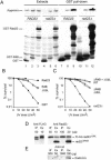
R4B is an autonomous Rad4-binding sequence that stabilizes Rad4, and restores significant UV resistance. (A) GST-fusion proteins (R4B or full-length Rad23) were expressed in yeast cells containing integrated Rad4–HA. Total extracts prepared from RAD23 and rad23Δ cells were analyzed by SDS–PAGE, and an immunoblot incubated with antibodies against GST (Extracts). The positions of GST, GST–R4B and GST–Rad23 are indicated by the bars on the left of the lower panel. Equal amounts of extracts were incubated with glutathione–Sepharose to purify GST and the GST-fusion proteins (GST pull-down). Note the lower levels of GST–R4B in extracts and on the GST beads. The same reactions were also tested for the presence of Rad4–HA (upper panel). Expression of GST–R4B restored normal levels of Rad4–HA in the extracts (lane 5). Furthermore, affinity-purified GST–R4B was associated with Rad4–HA at comparable levels as the full-length GST–Rad23 protein (compare lanes 8, 9 and 11, 12). The relatively lower levels of Rad4–HA in lanes 8 and 11 reflect the lower expression of the GST–R4B construct (see lower panel). (B) The ability of GST–R4B to bind Rad4-HA led us to examine its capacity to suppress the UV-sensitivity of rad23Δ. Both rad23ΔR4B and ΔUbLrad23 conferred an intermediate level of resistance. Wild-type, solid square; rad23Δ + GST, solid triangle; rad23Δ + GST–ΔUbLrad23, open triangle; rad23Δ + GST-rad23ΔR4B, open square. (C) To determine if rad23ΔR4B and ΔUbLrad23 functioned in distinct pathways, both proteins were simultaneously expressed in rad23Δ cells containing integrated Rad4–HA. Complete suppression of the UV-sensitivity of rad23Δ was detected. (Wild-type, solid square; rad23Δ, solid circle; rad23Δ + GST–ΔUbLrad23, open circle; rad23Δ + GST–rad23ΔR4B, open triangle; rad23Δ + GST–ΔUbLrad23 + GST–rad23ΔR4B, open diamond.) (D) Plasmids expressing ΔUbLrad23 and FLAG–rad23ΔR4B were transformed into cells expressing physiological levels of Rad4–HA. Protein extracts were prepared and incubated with antibodies against FLAG. The purified FLAG-rad23ΔUbL protein is shown in the left panel (lower band is antibody heavy chain). We examined 10 μg of total protein to detect FLAG-rad23ΔUbL, applied 50 μg of total protein to recover FLAG-rad23ΔUbL by immunoprecipitation. Immunoblots containing similar immunoprecipitation reactions were probed with antibodies against Rad23, so that both ΔUbLrad23 and FLAG–rad23ΔR4B would be detected (right panel). As expected, both proteins were readily detected in the extract lanes (Ex). Significantly, we detected no evidence for ΔUbLrad23/FLAG–rad23ΔR4B dimerization (see IP lanes). (We tested up to 1 mg of protein in the immunoprecipations, and did not detect an interaction.) (E) Immunoblots containing reactions with much larger quantity of protein extracts were reacted with antibodies against HA. We failed to detect Rad4–HA in association with FLAG–rad23ΔR4B, demonstrating the absence of dimerization.

Similar articles

Cited by

References

    1. Batty D.P. and Wood,R.D. (2000) Damage recognition in nucleotide excision repair of DNA2. Gene, 241, 193–204. - PubMed
    1. Prakash S. and Prakash,L. (2000) Nucleotide excision repair in yeast. Mutat. Res., 451, 13–24. - PubMed
    1. Lambertson D., Chen,L. and Madura,K. (1999) Pleiotropic defects caused by loss of the proteasome-interacting factors Rad23 and Rpn10 of Saccharomyces cerevisiae. Genetics, 153, 69–79. - PMC - PubMed
    1. Elsasser S., Chandler-Militello,D., Muller,B., Hanna,J. and Finley,D. (2004) Rad23 and Rpn10 serve as alternative ubiquitin receptors for the proteasome. J. Biol. Chem., 279, 26817–26822. - PubMed
    1. Schauber C., Chen,L., Tongaonkar,P., Vega,I., Lambertson,D., Potts,W. and Madura,K. (1998) Rad23 links DNA repair to the ubiquitin/proteasome pathway. Nature, 391, 715–718. - PubMed

Publication types

MeSH terms

Substances