. 2004 Dec 16;23(58):9438-46.
doi: 10.1038/sj.onc.1207988.

A gene highly expressed in tumor cells encodes novel structure proteins

Affiliations

A gene highly expressed in tumor cells encodes novel structure proteins

Nianli Sang et al. Oncogene. .

Abstract

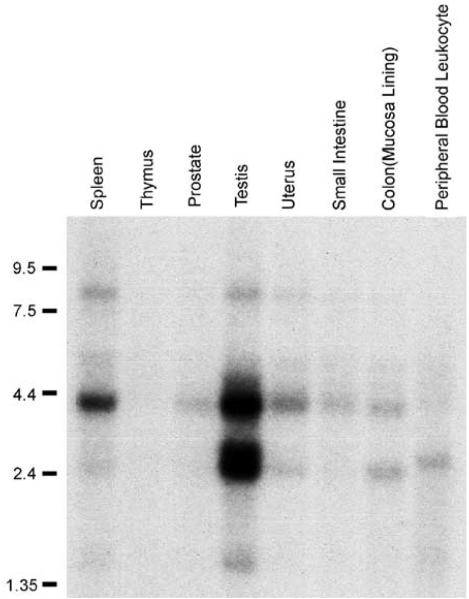
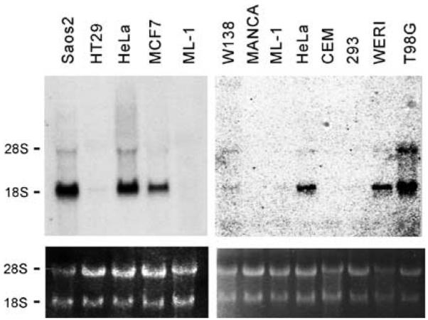
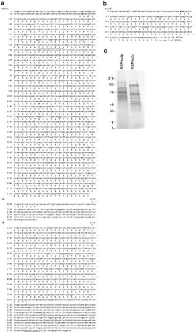
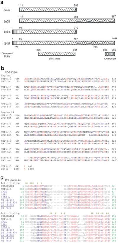
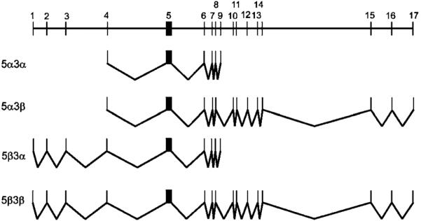
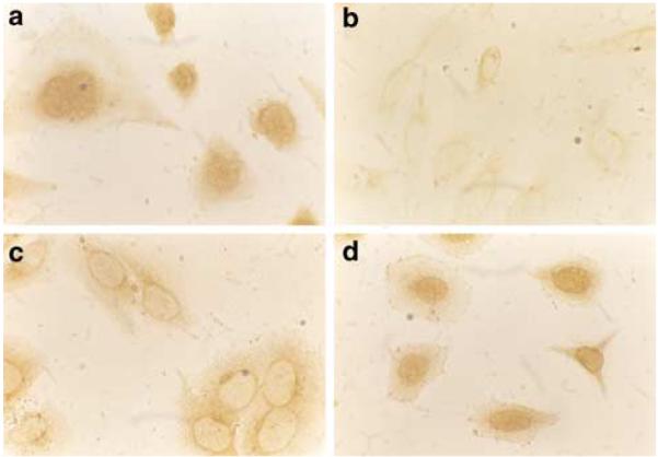
We isolated several related but distinct cDNA clones encoding novel structure proteins (NSP) when screening a cDNA library. Analysis revealed that these cDNAs and several similar ESTs in the public databases are derived from a single gene of 17 exons that span a minimum of 227-kb region. This gene is located at chromosome 17p11.2, a region frequently amplified in human gliomas and osteosarcomas, and involved in Birt-Hogg-Dube syndrome, a tumor-prone syndrome. The major coding sequences shared by all isolated transcripts are predicted to encode SMC (structural maintenance of chromosome)/SbcC ATPase motifs and coiled-coil domains commonly seen in motor or structure proteins. Two 5'-end and two 3'-end variants (type 5alpha/beta and 3alpha/beta, respectively) were identified, making a total of four possible transcripts. Both 5alpha and 5beta variants were detected in human testis mRNA, but only type 5alpha was detectable in RNA samples extracted from HeLa cells. The unique carboxyl-terminus of 3beta contains a Ca(2+)-dependent actin-binding domain. Immunohistochemistry studies revealed that NSPs were mostly localized to nuclei. Northern blot analysis demonstrated two major bands and the expression levels are tremendously high in testis while barely detectable in other normal tissues examined. Interestingly, NSP5alpha3alpha is highly expressed in some tumor cell lines. These results suggest that NSPs represent a new family of structure proteins with a possible role in nuclear dynamics during cell division, and that NSP5alpha3alpha may serve as a tumor marker.


Figures

Figure 1
Figure 1
Expression screening identifies cDNA fragments in HeLa cells. (a) Schematic illustration of several overlapping cDNA clones recognized by an anti-pRb antibody. (b) Northern blot shows the existence of more than one transcript in HeLa cells. Total RNA was extracted from HeLa cells and separated on denaturing agarose gel with molecular weight marker. Probe B indicated in (a) was used for the detection. (c) FISH analysis reveals that NSP is a single-copy gene localized at chromosome 17p12.2. Panels a and c show the FISH signals on chromosome 17; panels b and d indicate the same mitotic figure stained with DAPI to identify chromosome 17
Figure 2
Figure 2
Expression pattern of NSP in normal tissues. Multitissue Northern blots were purchased from Clontech (BD Science) and detected by Probe B indicated in Figure 1a
Figure 3
Figure 3
NSP5α3α is overexpressed in several tumor cell lines. Total RNA samples were extracted from the cell lines indicated. The samples were fractionated and transferred onto nylon membranes and hybridized with Probe B indicated in Figure 1a
Figure 4
Figure 4
Nucleotide and amino-acid sequences of NSPs. Putative translation start codons are in bold face. Putative nuclear localization signal is underlined. An alternative poly-A-tail site is underlined. (a) NSP5α3α and 5α3β. (b) Nucleotide and amino-acid sequences of the 5′-end variants from testis (NSP5α). (c) In vitro transcription and translation of NSP5α3α and NSP5α3β. SDS–PAGE shows that the longest translational products have the expected sizes. Faster migration species represent products translated from internal initiating codons
Figure 5
Figure 5
Structure analysis of NSP proteins. (a) Schematic illustration of conserved domains of NSP. Amino-acid numbering of conserved domains was based on NSP5α3β. (b) Alignment of the core homologous region of NSP with the conserved domain of SMC (KOG0161). (c) Alignment of the CH domains of NSP3β with other CH containing proteins. Surface required for actin binding is indicated by ‘#’
Figure 6
Figure 6
Genomic organization of NSP gene and the splicing schemes
Figure 7
Figure 7
Subcellular localization of NSP gene products. Saos-2 and HeLa cells were cultured in chamber slides and proceeded for immunohistochemistry studies as described in Material and methods. The crossreactive antibody recognized nuclear signals in the absence of pRb (a). (b) Normal mouse serum gave no signal. (c) XZ37 failed to stain the nuclei of Saos-2 cells (c), but detected pRb in HeLa cells (d)

References

    1. Allen DL, Leinwand LA. J. Biol. Chem. 2002;277:45323–45330. - PubMed
    1. Ample C, Vandekerckhove J. Semin. Cell Biol. 1994;5:175–182. - PubMed
    1. Banuelos S, Saraste M, Garugo KD. Structure. 1998;6:1419–1431. - PubMed
    1. Britton RA, Lin DC, Grossman AD. Genes Dev. 1998;12:1254–1259. - PMC - PubMed
    1. Carugo KD, Banuelos S, Saraste M. Nat. Struct. Biol. 1997;4:175–179. - PubMed

Publication types

Substances