. 2004 Dec 28;101(52):18129-34.
doi: 10.1073/pnas.0407817101. Epub 2004 Dec 16.

Functional assembly of engineered myocardium by electrical stimulation of cardiac myocytes cultured on scaffolds

Affiliations

Functional assembly of engineered myocardium by electrical stimulation of cardiac myocytes cultured on scaffolds

Milica Radisic et al. Proc Natl Acad Sci U S A. .

Abstract

The major challenge of tissue engineering is directing the cells to establish the physiological structure and function of the tissue being replaced across different hierarchical scales. To engineer myocardium, biophysical regulation of the cells needs to recapitulate multiple signals present in the native heart. We hypothesized that excitation-contraction coupling, critical for the development and function of a normal heart, determines the development and function of engineered myocardium. To induce synchronous contractions of cultured cardiac constructs, we applied electrical signals designed to mimic those in the native heart. Over only 8 days in vitro, electrical field stimulation induced cell alignment and coupling, increased the amplitude of synchronous construct contractions by a factor of 7, and resulted in a remarkable level of ultrastructural organization. Development of conductive and contractile properties of cardiac constructs was concurrent, with strong dependence on the initiation and duration of electrical stimulation.


Figures
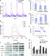
Fig. 1.
Fig. 1.
Functional properties of engineered cardiac constructs. (A) Contraction amplitude of paced constructs cultured for a total of 8 days, with or without electrical stimulation, shown as a fractional change in the construct surface area. (B) Contraction amplitude progressively increased with time. One representative contraction cycle is shown measured after 1, 2, 3, and 4 days of cultivation with electrical stimulation. (C and D) ET decreased (C) and maximum capture rate increased (D) significantly both with time in culture and because of electrical stimulation. (E) Electrical potentials recorded in the stimulated group after 8 days of culture resembled the action potentials measured in native heart ventricles. S, stimulus; R, response. (F) Protein levels determined by Western blots at the end of preculture (day 3) and for stimulated and nonstimulated constructs at the end of culture (day 8). CK-MM, creatine kinase-MM. (G) Relative protein amounts as determined from Western blots by using integrated pixel density analysis. * denotes statistically significant difference (P < 0.05; Tukey's post hoc test with one-way ANOVA, n = 5–10 samples per group and time point).
Fig. 2.
Fig. 2.
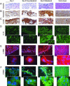
Histomorphology and expression of cardiac proteins. Representative sections of constructs at the end of preculture (day 3) and culture (day 8, with or without electrical stimulation); sections of native ventricles are shown for comparison. (A) Hematoxylin/eosin (H and E). (B) Tn-I (brown). (C) Sarcomeric α-actin (brown). Arrows denote myotubes. (D) Cx-43 (green). (E) α-MHC (red). Blue, cell nuclei; red arrows, positive cells; blue arrows, negative cells. (F) β-MHC (green). Blue, cell nuclei; yellow arrows, positive cells; blue arrows, negative cells; yellow arrows in Insets, striations. (Scale bars: AF, 50 μm; DF Insets, 20 μm.)
Fig. 3.
Fig. 3.
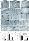
Ultrastructural organization. Shown are representative micrographs of stimulated and nonstimulated constructs after 8 days of cultivation, compared with neonatal rat ventricles. (A) Cell shape and orientation. (B) Overview of myofibrils. (C) Structure of a sarcomere. (D) Intercalated disk. (E) Gap junctions. (Insets) T-tubule. (F and G) Morphometric analysis. Volume fractions occupied by nuclei, sarcomeres, and mitochondria (F) and the frequency of membrane junctions (G). IC, intercalated. * denotes statistically significant differences between the groups (P < 0.05; Tukey's post hoc test with one-way ANOVA; n = 18–46 samples per group and time point). (Scale bars: A and B, 2 μm; C and D, 1 μm; E and Insets, 0.5 μm.)
Fig. 4.
Fig. 4.
Concurrent and progressive development of conductive and contractile properties of cardiac constructs cultured in vitro. During phase 1 (preculture without electrical stimulation), cells accumulate and assemble conductive and contractile proteins lost or disorganized during isolation from heart tissue; electrical stimulation has an inhibitory role. During phase 2 (cultivation with electrical stimulation), constructs are cultured with the application of electrical stimulation. The cells oriented in the direction of electrical field will be the first ones to elongate and establish gap junctions with neighboring cells. As the contractions begin, they drive the organization of sarcomeres and thereby increase the contractile force in response to electrical stimuli. Electrical stimulation enhances the development of ultrastructural and contractile properties of the individual cells (arrow to the right) and increases the number of functionally coupled cells engaged in synchronous contractions of the constructs (arrow to the left). Reproduced with permission from illustrator Jennifer Fairman (Copyright 2004, Fairman Studios).

References

    1. American Heart Association (2003) Heart Disease and Stroke Statistics: 2004 Update (Am. Heart Assoc., Dallas).
    1. Carrier, R. L., Papadaki, M., Rupnick, M., Schoen, F. J., Bursac, N., Langer, R., Freed, L. E. & Vunjak-Novakovic, G. (1999) Biotechnol. Bioeng. 64, 580–589. - PubMed
    1. Eschenhagen, T., Fink, C., Remmers, U., Scholz, H., Wattchow, J., Woil, J., Zimmermann, W., Dohmen, H. H., Schafer, H., Bishopric, N., et al. (1997) FASEB J. 11, 683–694. - PubMed
    1. Leor, J., Aboulafia-Etzion, S., Dar, A., Shapiro, L., Barbash, I. M., Battler, A., Granot, Y. & Cohen, S. (2000) Circulation 102, Suppl. 3, III56–III61. - PubMed
    1. Li, R.-K., Jia, Z. Q., Weisel, R. D., Mickle, D. A. G., Choi, A. & Yau, T. M. (1999) Circulation 100, Suppl., II63–II69. - PubMed

Publication types

MeSH terms

LinkOut - more resources