

Mannose binding lectin and C3 act as recognition molecules for infectious agents in the vagina
- PMID: 15606621
- PMCID: PMC1809267
- DOI: 10.1111/j.1365-2249.2005.02660.x
Mannose binding lectin and C3 act as recognition molecules for infectious agents in the vagina
Abstract
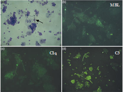
In our study we examined the early complement components in patients with bacterial vaginosis (BV), vulvovaginal candidiasis (VVC) and in healthy controls. The levels of C1q, mannose-binding lectin (MBL) and C3 were measured by ELISA in the cervicovaginal lavage (CVL) from gynaecological patients and controls. No significant differences were observed in the levels of these proteins in the three study groups. Immunofluorescence analysis of the clue cells and Candida hyphae from BV and VVC patients for surface-bound complement components showed the presence of C3, while C1q was undetectable. MBL was revealed on clue cells but not on Candida. Binding of MBL to Candida, grown or cytocentrifuged from the CVL of VVC patients, was found to be pH dependent and occurred between pH 4.5 and pH 5.5. In conclusion, we demonstrated that MBL and C3 present in the vaginal cavity act as recognition molecules for infectious agents that colonize the cervicovaginal mucosa. Our finding that MBL, but not C1q, binds to bacteria and fungi in vagina suggests that the lectin and classical pathways of complement activation may play a different role in immune defence in the female genital tract.
Figures


Similar articles
-
Mannose-binding lectin and vulvovaginal candidiasis.Int J Gynaecol Obstet. 2006 Jan;92(1):43-7. doi: 10.1016/j.ijgo.2005.08.024. Epub 2005 Oct 26. Int J Gynaecol Obstet. 2006. PMID: 16256117
-
Mannose-binding lectin is produced by vaginal epithelial cells and its level in the vaginal fluid is influenced by progesterone.Mol Immunol. 2010 Nov-Dec;48(1-3):281-6. doi: 10.1016/j.molimm.2010.07.016. Epub 2010 Aug 21. Mol Immunol. 2010. PMID: 20728220
-
Leishmania (Viannia) braziliensis: interaction of mannose-binding lectin with surface glycoconjugates and complement activation. An antibody-independent defence mechanism.Parasite Immunol. 2005 Sep;27(9):333-40. doi: 10.1111/j.1365-3024.2005.00782.x. Parasite Immunol. 2005. PMID: 16149991
-
Mannan binding lectin and its interaction with immunoglobulins in health and in disease.Immunol Lett. 2006 Aug 15;106(2):103-10. doi: 10.1016/j.imlet.2006.05.007. Epub 2006 Jun 12. Immunol Lett. 2006. PMID: 16814399 Review.
-
Mannose-binding lectin (MBL) and vascular complications in diabetes.Horm Metab Res. 2005 Apr;37 Suppl 1:95-8. doi: 10.1055/s-2005-861372. Horm Metab Res. 2005. PMID: 15918118 Review.
Cited by
-
The Interaction of Human Pathogenic Fungi With C-Type Lectin Receptors.Front Immunol. 2018 Jun 4;9:1261. doi: 10.3389/fimmu.2018.01261. eCollection 2018. Front Immunol. 2018. PMID: 29915598 Free PMC article. Review.
-
Immunopathology of Recurrent Vulvovaginal Infections: New Aspects and Research Directions.Front Immunol. 2019 Aug 28;10:2034. doi: 10.3389/fimmu.2019.02034. eCollection 2019. Front Immunol. 2019. PMID: 31555269 Free PMC article. Review.
-
The complement system at the embryo implantation site: friend or foe?Front Immunol. 2012 Mar 19;3:55. doi: 10.3389/fimmu.2012.00055. eCollection 2012. Front Immunol. 2012. PMID: 22566936 Free PMC article.
-
Mannan-binding lectin regulates dendritic cell maturation and cytokine production induced by lipopolysaccharide.BMC Immunol. 2011 Jan 1;12:1. doi: 10.1186/1471-2172-12-1. BMC Immunol. 2011. PMID: 21194488 Free PMC article.
-
Vaginal delivery of vaccines.Adv Drug Deliv Rev. 2021 Nov;178:113956. doi: 10.1016/j.addr.2021.113956. Epub 2021 Sep 1. Adv Drug Deliv Rev. 2021. PMID: 34481031 Free PMC article. Review.
References
-
- Anon. Non-reported sexually transmitted disease. Morb Mortal Wkly Rep. 1979;28:61–2.
-
- Cauci S, Monte R, Driussi S, Lanzafame P, Quadrifoglio F. Impairment of the mucosal immune system: IgA and IgM cleavage detected in vaginal washings of a subgroup of patients with bacterial vaginosis. J Infect Dis. 1998;178:1698–706. - PubMed
-
- Amsel R, Totten PA, Spiegel CA, Chen KC, Eschenbach D, Holmes KK. Nonspecific vaginitis. Diagnostic criteria and microbial and epidemiologic associations. Am J Med. 1983;74:14–22. - PubMed
-
- Mestecky J, Fultz PN. Mucosal immune system of the human genital tract. J Infect Dis. 1999;179(Suppl. 3):S470–4. - PubMed
-
- Volanakis JE. Overview of the Complement System. In: Frank MM, editor. The Human Complement System in Human and Disease. New York: Marcel Dekker; 1998. pp. 9–32.
Publication types
MeSH terms
Substances
LinkOut - more resources
Full Text Sources
Other Literature Sources
Miscellaneous
