. 2005 Jan;114(1):53-62.
doi: 10.1111/j.1365-2567.2004.02005.x.

The role of chemokines and extracellular matrix components in the migration of T lymphocytes into three-dimensional substrata

Affiliations

The role of chemokines and extracellular matrix components in the migration of T lymphocytes into three-dimensional substrata

Jyrki Ivanoff et al. Immunology. 2005 Jan.

Abstract

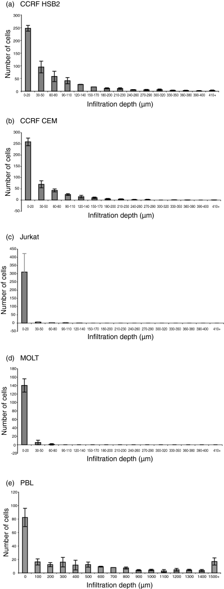
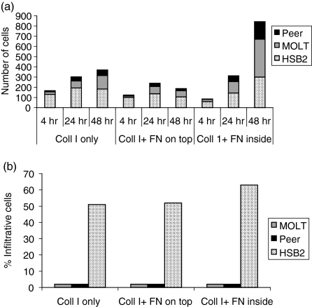
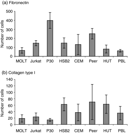
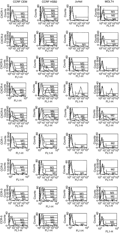
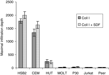
The role of chemokines and their interactions with extracellular matrix components (ECM) or the capacity of T cells to migrate into and accumulate within three-dimensional (3D) collagen type 1 substrata was studied. We examined the influence of chemokines and fibronectin on the infiltration properties of non-infiltrative (do not migrate into 3D substrata) and spontaneously infiltrative (migrate into 3D substrata) T-cell lines. Infiltrative and non-infiltrative T-acute lymphocytic leukaemic cell lines exhibited no consistent differences with respect to the expression of various chemokine receptors or beta(1)-integrins. Chemokines presented inside the collagen increased the depth of migration of infiltrative T-cell lines, but did not render non-infiltrative T-cell lines infiltrative, although they augmented the attachment of non-infiltrative T-cell lines to the upper surface of the collagen. The presence of fibronectin inside the collagen did not render non-infiltrative T-cell lines infiltrative, but markedly augmented the migration of 'infiltrative' T-cell lines into collagen. Both infiltrative and non-infiltrative T-cell lines showed migratory responses to chemokines in Boyden assays (migration detected on 2D substrata). These results indicate that the process of T-cell infiltration/migration into 3D substrata depends on a tissue penetration mechanism distinguishable from migration on 2D substrata and that the basic capacity of T cells to infiltrate is independent of chemokines and ECM components applied as attractants.


Figures

Figure 1
Figure 1
Infiltration capacity distinguishes separate T-cell lines. The figure shows a comparison of the infiltration of the leukaemic T-cell lines CCRF HSB2, CCRF CEM, Jurkat, MOLT4 and normal blood T lymphocytes [peripheral blood lymphocytes (PBL): 92–100% of the PBL cells attaching the collagen were identified as CD3 positive by immunocytochemistry] into collagen type I gels. For each cell line, adhesion and infiltration into the collagen were determined at 10-µm intervals (PBL at 100-µm intervals) by the use of an inverted microscope (magnification × 200) and a digital depth meter. The cell lines CCRF HSB2 and CCRF CEM infiltrated > 410 µm (leading front) over a 24-hr time-period (a) and (b), while Jurkat infiltrated to a minor extent, or not at all, during the same period (c). The cell line MOLT4 (d) showed no infiltration into the collagen, although the cells attached to the upper surface of the collagen to the same extent as the infiltrating cell lines. Normal T cells showed a pronounced capacity to infiltrate collagen (e). The figure shows mean values ± standard error of the mean (SEM) (expressed as mean number of cells per cm2) of seven positions after 24 hr in three single wells.
Figure 2
Figure 2
The influence of fibronectin (FN), present inside and on top of collagen type I gels (Coll I), on T-cell attachment to the gel (a) and on infiltration into the gel (b). The attachment refers to all cells throughout the collagen per microscope field (× 200) (a), and infiltrative cells refer to the percentage of cells migrating > 20 µm into the collagen gel (b). The presence of fibronectin in the collagen did not render the non-infiltrative T-cell lines Peer and MOLT infiltrative, although it increased the number of cells on top of the collagen. In contrast, fibronectin present inside the collagen markedly increased the number of cells migrating into the collagen in the infiltrative T-cell line, CCRF HSB2. The results shown in this figure demonstrate that an adhesive component inside the collagen does not change the infiltrative behaviour of the cells. The figure shows the results of one representative experiment.
Figure 3
Figure 3
Migration of T-cell lines and peripheral blood lymphocytes (PBL) through Boyden filters coated on the lower side with fibronectin (10 µg/ml) (a) or with collagen type I (40 µg/ml) (b). The Boyden assays detected no consistent differences in migration capacity between infiltrative (CCRF HSB2, CCRF CEM) and non-infiltrative (MOLT, Jurkat, P30) T cells. Furthermore, the T-cell line Peer migrated on both collagen and fibronectin, despite its failure to infiltrate. Normal PBL, which showed pronounced infiltration into collagen (Fig. 1e), showed the same, or a somewhat lower, magnitude of hapotactic migration than the T-cell lines. The number of migrating cells represents the mean number ± standard error of the mean (SEM) per field of six microscopic fields (magnification × 400).
Figure 4
Figure 4
Chemokine receptor expression does not differ between infiltrating and non-infiltrating T cells. The figure shows flow cytometry analysis of the expression of chemokine receptors in two infiltrative (CCRF HSB2, CCRF CEM) and two non-infiltrative (MOLT4, Jurkat) T-leukaemia cell lines. A high percentage of the T-cell lines were CXCR-4, CCR-3 and CXCR-5 positive. The T-cell lines showed a variable expression of CXCR-1, CXCR-2, CXCR-3 and CCR-5, and a low expression of CCR-1, CCR-2 and CCR-6 (data not shown). The symbols M1-4 denote different comparisons of the area under the curves of the immunoglobulin G (IgG) control and the monoclonal antibodies to the chemokines, respectively.
Figure 5
Figure 5
Chemokines stimulate adhesion and migration, but do not render non-infiltrative T cells infiltrative. The figure shows the influence of stromal-derived factor-1α (SDF-1α) (50 ng/ml) on the adhesion and infiltration on/into collagen type I gels of infiltrative (CCRF HSB2, CCRF CEM) (a) and (b) and non-infiltrative (Jurkat, MOLT4) (c) and (d) T-cell lines. The chemokine was presented to the T cells in different ways: –/–, no chemokines; +/–, chemokines added in the cell culture medium; –/+, chemokines added in the collagen type I gel; and +/+, chemokines added both in the medium and in the gel. Adhesion to and infiltration into collagen was determined in three experiments (in seven positions per experiment) at every 10 µm throughout the gel (magnification × 200), and the figure shows the mean values ± standard error of the mean (SEM). SDF-1α increased the number of cells infiltrating > 60 µm into collagen of ‘infiltrative’ T-cell lines (CCRF HSB2, P < 0·05; CCRF CEM, P < 0·001) when presented in the gel, but the chemokine had no effect on infiltration when present with the cells (a) and (b). In contrast, SDF-1α exerted a significant stimulatory effect on the adhesion of non-infiltrative T-cell lines to the upper surface, comprising > 50 µm of the collagen, when present with the cells (MOLT4, P < 0·05) or in the gel (Jurkat, P < 0·05; MOLT4, P < 0·05), but the infiltrating capacity of the cells was not affected (c) and (d).
Figure 6
Figure 6
The influence of stromal-derived factor-1α (SDF-1α) on the maximal infiltration depth of infiltrative and non-infiltrative T-cell lines when the chemokine was presented on the opposite side of the collagen layer at a concentration of 50 ng/ml. SDF-1α only marginally increased the infiltration of infiltrative T cells (CCRF HSB2, CCRF CEM, HUT), and had no stimulatory effect on the infiltration of non-infiltrative T-cell lines (MOLT, P30, Jurkat, Peer). The figure shows the mean maximal infiltration depth ± standard error of the mean (SEM) determined from 10 microscopic fields per cell line (× 100) of cells allowed to migrate for 24 hr.

References

    1. Alon R, Rossiter H, Wang X, Springer TA, Kupper TS. Distinct cell surface ligands mediate T lymphocyte attachment and rolling on P and E selectin under physiological flow. J Cell Biol. 1994;127:1485–95. - PMC - PubMed
    1. Hauzenberger D, Klominek J, Bergström SE, Sundqvist KG. T lymphocyte migration: the influence of interactions via adhesion molecules, the T cell receptor and cytokines. Crit Rev Immunol. 1995;15:285–316. - PubMed
    1. Bird IN, Spragg JH, Ager A, Matthews N. Studies of lymphocyte transendothelial migration: analysis of migrated cell phenotypes with regard to CD31 (PECAM-1), CD45RA and CD45RO. Immunology. 1993;80:553–60. - PMC - PubMed
    1. Lawrence MB, Springer TA. Leukocytes roll on a selectin at physiologic flow rates: distinction from and prerequisite for adhesion through integrins. Cell. 1991;65:859–73. - PubMed
    1. Brenner B, Gulbins E, Schlottmann K, et al. l-Selectin activates the Ras pathway via the tyrosine kinase p56lck. Proc Natl Acad Sci USA. 1996;93:15376–81. - PMC - PubMed

Publication types

LinkOut - more resources