. 2004 Dec 28;101(52):18111-6.
doi: 10.1073/pnas.0408558102. Epub 2004 Dec 17.

Heterozygosity for Lmna deficiency eliminates the progeria-like phenotypes in Zmpste24-deficient mice

Affiliations

Heterozygosity for Lmna deficiency eliminates the progeria-like phenotypes in Zmpste24-deficient mice

Loren G Fong et al. Proc Natl Acad Sci U S A. .

Abstract

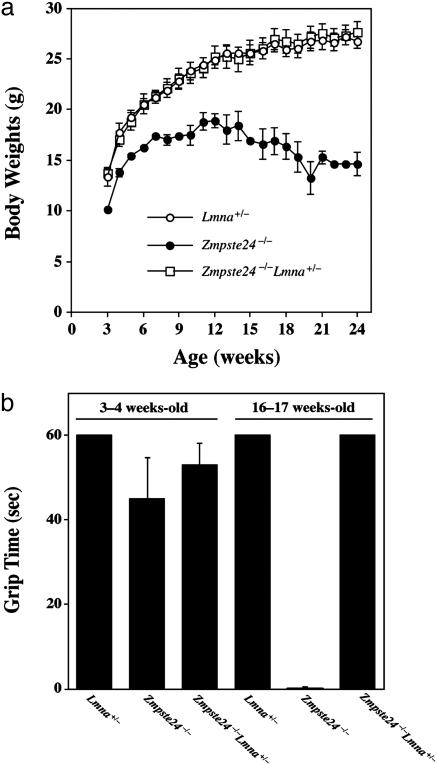
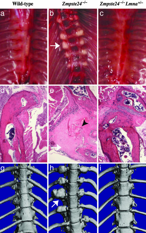
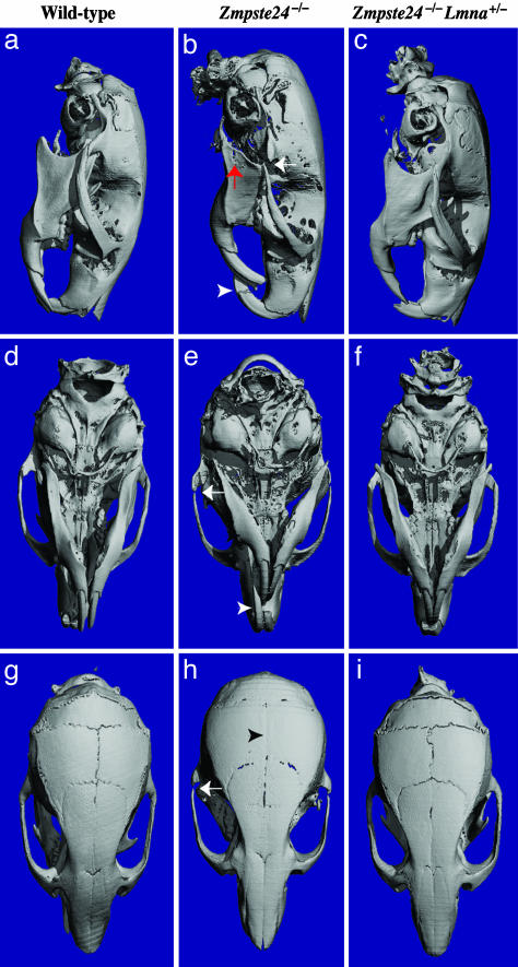
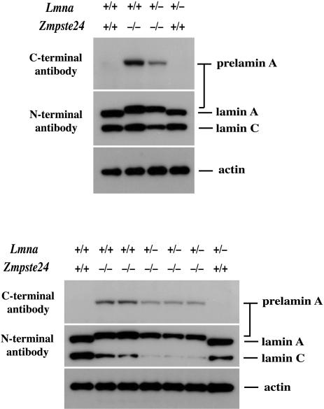
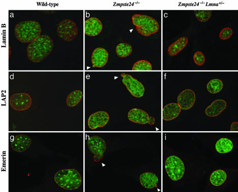
Zmpste24 is a metalloproteinase required for the processing of prelamin A to lamin A, a structural component of the nuclear lamina. Zmpste24 deficiency results in the accumulation of prelamin A within cells, a complete loss of mature lamin A, and misshapen nuclear envelopes. Zmpste24-deficient (Zmpste24(-/-)) mice exhibit retarded growth, alopecia, micrognathia, dental abnormalities, osteolytic lesions in bones, and osteoporosis, which are phenotypes shared with Hutchinson-Gilford progeria syndrome, a human disease caused by the synthesis of a mutant prelamin A that cannot undergo processing to lamin A. Zmpste24(-/-) mice also develop muscle weakness. We hypothesized that prelamin A might be toxic and that its accumulation in Zmpste24(-/-) mice is responsible for all of the disease phenotypes. We further hypothesized that Zmpste24(-/-) mice with half-normal levels of prelamin A (Zmpste24(-/-) mice with one Lmna knockout allele) would be subjected to less toxicity and be protected from disease. Thus, we bred and analyzed Zmpste24(-/-)Lmna(+/-) mice. As expected, prelamin A levels in Zmpste24(-/-)Lmna(+/-) cells were significantly reduced. Zmpste24(-/-)Lmna(+/-) mice were entirely normal, lacking all disease phenotypes, and misshapen nuclei were less frequent in Zmpste24(-/-)Lmna(+/-) cells than in Zmpste24(-/-) cells. These data suggest that prelamin A is toxic and that reducing its levels by as little as 50% provides striking protection from disease.


Figures

Fig. 1.
Fig. 1.
Growth rates and grip strength in mice. (a) Female Zmpste24–/– Lmna+/– mice (n = 6) exhibit normal growth, indistinguishable from wild-type mice (n = 5). Similar results were obtained with male mice (data not shown). (b) Muscle strength in 3- to 4-week old and 16- to 17-week-old female wild-type (Lmna+/–, n = 7), Zmpste24–/– (n = 5), and Zmpste24–/–Lmna+/– mice (n = 9), as judged by the length of time that they were able to hang on to an upside-down grid (1) (grip time; P < 0.001). Similar results were observed for male mice. Differences became statistically significant at 8 weeks of age.
Fig. 2.
Fig. 2.
Fractures of ribs near the costovertebral joint occur in Zmpste24–/– mice but not in wild-type or Zmpste24–/–Lmna+/– mice. (ac) Photographs of the thorax after removal of the heart and lungs, revealing fracture callus in a Zmpste24–/– mouse (b, arrow indicating callus around a rib fracture). (df) Hematoxylin/eosin-stained sections of ribs near the costovertebral joint. In the Zmpste24–/– mouse (e), fibrotic fracture callus (white arrow) contains fragments of necrotic bone (black arrowhead). Identical results were observed in three mice of each genotype. (gi) Surface renderings of μCT scans of the thoracic spine in 28-week-old wild-type, Zmpste24–/–, and Zmpste24–/–Lmna+/– mice. In the Zmpste24–/– mouse (h), fracture callus is indicated by a white arrow.
Fig. 3.
Fig. 3.
Absence of skull abnormalities in Zmpste24–/–Lmna+/– mice at 20 weeks of age, as judged by surface renderings of μCT scans. (a, d, and g) Wild-type mouse. (b, e, and h) Zmpste24–/– mouse. (c, f, and i) Zmpste24–/–Lmna+/– mouse. Wild-type and Zmpste24–/–Lmna+/– skulls were indistinguishable. Skulls of Zmpste24–/– mice exhibited a smaller mandible, in this case with an osteolytic lesion (red arrow in b); elongated incisors (white arrowhead in b and e); osteolytic lesion in the posterior portion of the zygomatic arch (white arrows in b, e, and h); and loss of the zigzag appearance of the cranial sutures (black arrowhead in H). Identical results were observed in two mice of each genotype.
Fig. 4.
Fig. 4.
Western blots of extracts from wild-type, Zmpste24–/–, and Zmpste24–/–Lmna+/– MEFs with a carboxyl (C)-terminal prelamin A antibody and an amino (N)-terminal lamin A/C antibody. Analyses from two independent experiments, with cells prepared from two different sets of embryos, are shown. Protein loading was assessed with an antibody against β-actin. Densitometry showed a 58.4 ± 4.1% reduction in prelamin A and a 78 ± 4.9% decrease in lamin C in the Zmpste24–/–Lmna+/– cells (n = 4) relative to the Zmpste24–/– cells.
Fig. 5.
Fig. 5.
Analysis of nuclear shape in wild-type, Zmpste24–/–, and Zmpste24–/–Lmna+/– MEFs by laser-scanning fluorescence microscopy. The nuclear envelope was visualized with antibodies to lamin B (ac), LAP2 (df), or emerin (gi) (red), and DNA was visualized with SYTOX Green staining (green). White arrowheads indicate blebs.

References

    1. Bergo, M. O., Gavino, B., Ross, J., Schmidt, W. K., Hong, C., Kendall, L. V., Mohr, A., Meta, M., Genant, H., Jiang, Y., et al. (2002) Proc. Natl. Acad. Sci. USA 99, 13049–13054. - PMC - PubMed
    1. Pendás, A. M., Zhou, Z., Cadiñanos, J., Freije, J. M. P., Wang, J., Hultenby, K., Astudillo, A., Wernerson, A., Rodríguez, F., Tryggvason, K. & Lopéz-Otín, C. (2002) Nat. Genet. 31, 94–99. - PubMed
    1. Tam, A., Nouvet, F. J., Fujimura-Kamada, K., Slunt, H., Sisodia, S. S. & Michaelis, S. (1998) J. Cell Biol. 142, 635–649. - PMC - PubMed
    1. Boyartchuk, V. L. & Rine, J. (1998) Genetics 150, 95–101. - PMC - PubMed
    1. Sullivan, T., Escalante-Alcalde, D., Bhatt, H., Anver, M., Bhat, N., Nagashima, K., Stewart, C. L. & Burke, B. (1999) J. Cell Biol. 147, 913–919. - PMC - PubMed

Publication types

MeSH terms