

doi: 10.1073/pnas.0408319102.
Epub 2004 Dec 17.
Surface-enhanced Raman scattering on tunable plasmonic nanoparticle substrates
Affiliations
- PMID: 15608058
- PMCID: PMC539806
- DOI: 10.1073/pnas.0408319102
Item in Clipboard
Surface-enhanced Raman scattering on tunable plasmonic nanoparticle substrates
Proc Natl Acad Sci U S A.
.
Abstract
Au and Ag nanoshells are investigated as substrates for surface-enhanced Raman scattering (SERS). We find that SERS enhancements on nanoshell films are dramatically different from those observed on colloidal aggregates, specifically that the Raman enhancement follows the plasmon resonance of the individual nanoparticles. Comparative finite difference time domain calculations of fields at the surface of smooth and roughened nanoshells reveal that surface roughness contributes only slightly to the total enhancement. SERS enhancements as large as 2.5 x 10(10) on Ag nanoshell films for the nonresonant molecule p-mercaptoaniline are measured.
Figures
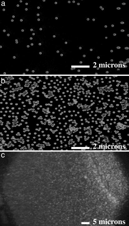
Representative environmental scanning electron microscope images of PVP/Au nanoshell films, characterized by the number of nanoshells per 2-μm spot (NS/spot). (a) 2.58 ± 0.32 NS/spot. (b) 16.66 ± 1.9 NS/spot. (c) Optical micrograph of a dense multilayer nanoshell film.
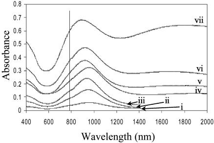
Absorption spectrum of the PVP/Au nanoshell films for each nanoshell density listed in Table 1. The pump laser wavelength of 782 nm is shown.
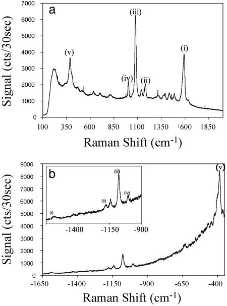
Stokes (a) and anti-Stokes (b) SERS spectrum of pMA on nanoshell film substrates. The 1,590-cm–1 (i), 1,180-cm–1 (ii), 1,077-cm–1 (iii), 1,003-cm–1 (iv), and 390-cm–1 (v) ring vibrational modes of pMA are indicated.
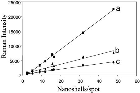
The 1,077-cm–1 (a), 1,590-cm–1 (b), and 390-cm–1 (c) Raman modes as a function of Au nanoshell density on the substrate.
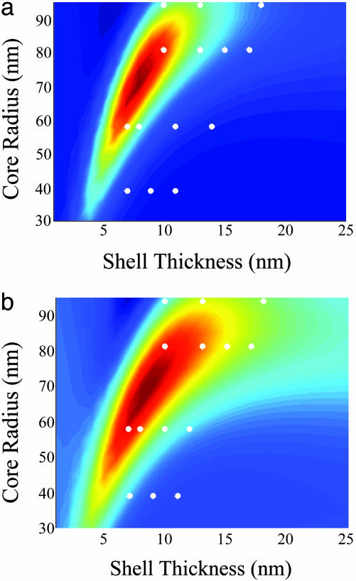
Calculated |Eshell(ωo)|2|ERaman(ωs)|2 of 1,590-cm–1 (a) and 390-cm–1 (b) modes as a function of silica core radius and silver shell thickness. The white dots indicate the fabricated nanoshell films.
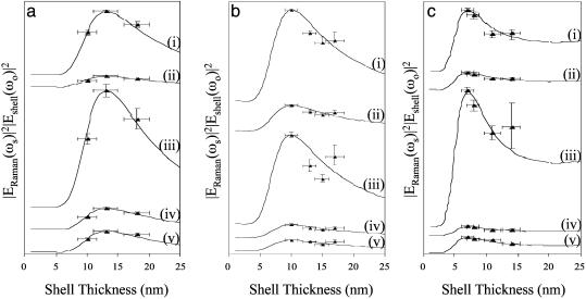
Comparison of the measured Raman modes to theoretical calculations extracted from the contour plots shown in Fig. 5. The normalized |Eshell(ωo)|2|ERaman(ωs)|2 of the 1,590-cm–1 (i), 1,180-cm–1 (ii), 1,077-cm–1 (iii), 1,003-cm–1 (iv), and 390-cm–1 (v) Stokes modes are plotted for each fabricated core radius, where a is 94 nm, b is 81 nm, and c is 58 nm.
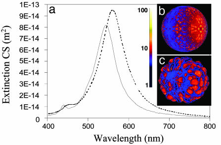
Finite-difference time-domain calculations of the far- and near-field plasmon response of a smooth and a rough but continuous nanoshell. (a) The extinction cross section of a smooth (solid line) and rough (dashed line) silver nanoshell with a 39-nm-radius core and a 9-nm-thick shell. The magnitudes of the electromagnetic field on the smooth nanoshell at the peak dipole resonance (545 nm) (b) and the rough nanoshell at the peak dipole resonance (562 nm) (c) are shown.
References
-
- Jeanmarie, D. L. & Van Duyne, R. P. (1977) J. Electroanal. Chem. 84, 1.
-
- Moskovits, M. (1985) Rev. Mod. Phys. 57, 783–826.
-
- Fleischmann, M., Hendra, P. & McMillan, A. (1974) Chem. Phys. Lett. 26, 163–166.
-
- Fleischmann, M., Hendra, P. J. & McQuillan, A. J. (1973) J. Chem. Soc. Chem. Commun. 3, 80–81.
-
- Maher, R. C., Cohen, L. F., Etchegoin, P., Hartigan, H. J. N., Brown, R. J. C. & Milton, M. J. T. (2004) J. Chem. Phys. 120, 11746–11753. - PubMed
Publication types
MeSH terms
Substances
LinkOut - more resources
Full Text Sources
Other Literature Sources
Miscellaneous
