

Comparison between TRF2 and TRF1 of their telomeric DNA-bound structures and DNA-binding activities
- PMID: 15608118
- PMCID: PMC2253311
- DOI: 10.1110/ps.04983705
Comparison between TRF2 and TRF1 of their telomeric DNA-bound structures and DNA-binding activities
Abstract
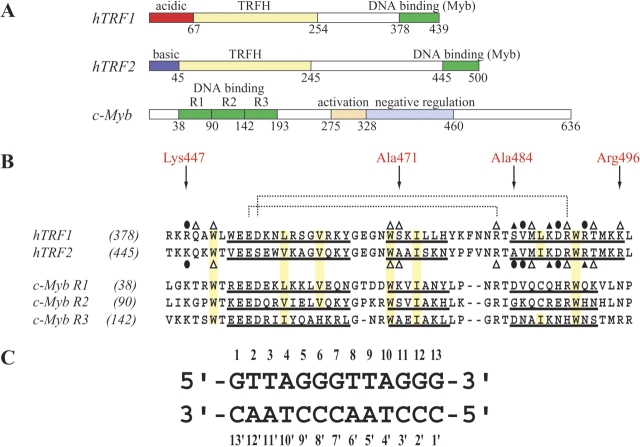
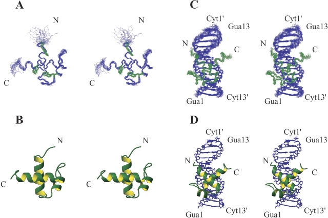
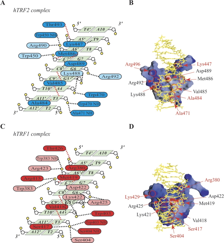
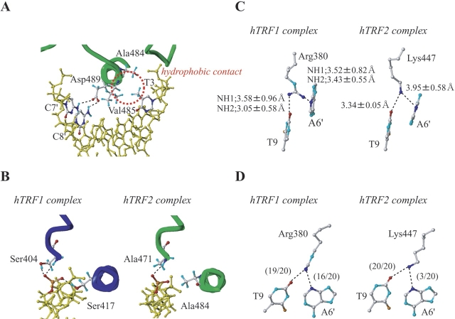
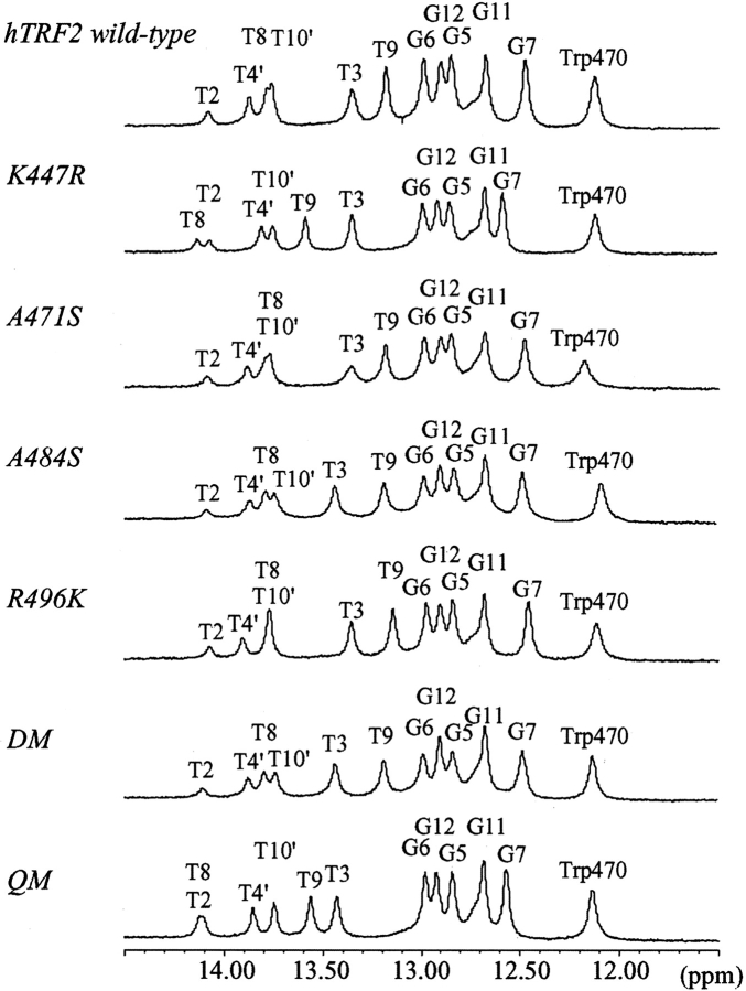
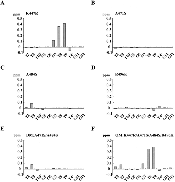
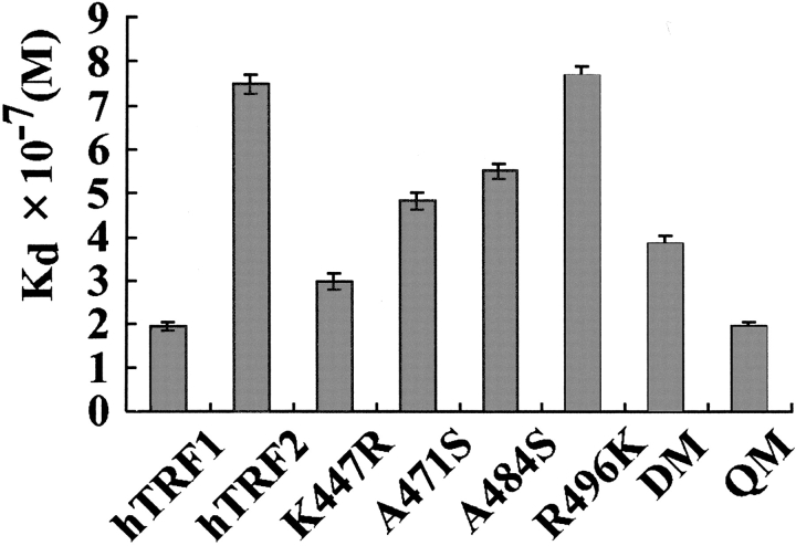
Mammalian telomeres consist of long tandem arrays of double-stranded telomeric TTAGGG repeats packaged by the telomeric DNA-binding proteins TRF1 and TRF2. Both contain a similar C-terminal Myb domain that mediates sequence-specific binding to telomeric DNA. In a DNA complex of TRF1, only the single Myb-like domain consisting of three helices can bind specifically to double-stranded telomeric DNA. TRF2 also binds to double-stranded telomeric DNA. Although the DNA binding mode of TRF2 is likely identical to that of TRF1, TRF2 plays an important role in the t-loop formation that protects the ends of telomeres. Here, to clarify the details of the double-stranded telomeric DNA-binding modes of TRF1 and TRF2, we determined the solution structure of the DNA-binding domain of human TRF2 bound to telomeric DNA; it consists of three helices, and like TRF1, the third helix recognizes TAGGG sequence in the major groove of DNA with the N-terminal arm locating in the minor groove. However, small but significant differences are observed; in contrast to the minor groove recognition of TRF1, in which an arginine residue recognizes the TT sequence, a lysine residue of TRF2 interacts with the TT part. We examined the telomeric DNA-binding activities of both DNA-binding domains of TRF1 and TRF2 and found that TRF1 binds more strongly than TRF2. Based on the structural differences of both domains, we created several mutants of the DNA-binding domain of TRF2 with stronger binding activities compared to the wild-type TRF2.
Figures
Similar articles
-
Solution structure of a telomeric DNA complex of human TRF1.Structure. 2001 Dec;9(12):1237-51. doi: 10.1016/s0969-2126(01)00688-8. Structure. 2001. PMID: 11738049
-
How the human telomeric proteins TRF1 and TRF2 recognize telomeric DNA: a view from high-resolution crystal structures.EMBO Rep. 2005 Jan;6(1):39-45. doi: 10.1038/sj.embor.7400314. EMBO Rep. 2005. PMID: 15608617 Free PMC article.
-
TRF1 and TRF2 binding to telomeres is modulated by nucleosomal organization.Nucleic Acids Res. 2015 Jul 13;43(12):5824-37. doi: 10.1093/nar/gkv507. Epub 2015 May 20. Nucleic Acids Res. 2015. PMID: 25999344 Free PMC article.
-
Post-translational modifications of TRF1 and TRF2 and their roles in telomere maintenance.Mech Ageing Dev. 2012 Jun;133(6):421-34. doi: 10.1016/j.mad.2012.05.002. Epub 2012 May 23. Mech Ageing Dev. 2012. PMID: 22634377 Review.
-
Telomere Repeat-Binding Factor 2 Is Responsible for the Telomere Attachment to the Nuclear Membrane.Adv Protein Chem Struct Biol. 2015;101:67-96. doi: 10.1016/bs.apcsb.2015.06.009. Epub 2015 Oct 21. Adv Protein Chem Struct Biol. 2015. PMID: 26572976 Review.
Cited by
-
One identity or more for telomeres?Front Oncol. 2013 Mar 15;3:48. doi: 10.3389/fonc.2013.00048. eCollection 2013. Front Oncol. 2013. PMID: 23509004 Free PMC article.
-
Telomerase and telomere-associated proteins: structural insights into mechanism and evolution.Structure. 2012 Jan 11;20(1):28-39. doi: 10.1016/j.str.2011.10.017. Structure. 2012. PMID: 22244753 Free PMC article. Review.
-
ERK1/2/MAPK pathway-dependent regulation of the telomeric factor TRF2.Oncotarget. 2016 Jul 19;7(29):46615-46627. doi: 10.18632/oncotarget.10316. Oncotarget. 2016. PMID: 27366950 Free PMC article.
-
Structural biology of telomeres and telomerase.Cell Mol Life Sci. 2020 Jan;77(1):61-79. doi: 10.1007/s00018-019-03369-x. Epub 2019 Nov 14. Cell Mol Life Sci. 2020. PMID: 31728577 Free PMC article. Review.
-
CGGBP1 phosphorylation constitutes a telomere-protection signal.Cell Cycle. 2014;13(1):96-105. doi: 10.4161/cc.26813. Epub 2013 Oct 23. Cell Cycle. 2014. PMID: 24196442 Free PMC article.
References
-
- Biedenkapp, H., Borgmeyer, U., Sippel, A.E., and Klempnauer, K.H. 1988. Viral myb oncogene encodes a sequence-specific DNA-binding activity. Nature 335 835–837. - PubMed
-
- Bilaud, T., Brun, C., Ancelin, K., Koering, C.E., Laroche, T., and Gilson, E. 1997. Telomeric localization of TRF2, a novel human telobox protein. Nat. Genet. 17 236–239. - PubMed
Publication types
MeSH terms
Substances
Associated data
- Actions
- Actions
LinkOut - more resources
Full Text Sources
Other Literature Sources
Research Materials
Miscellaneous
