

The CATH Domain Structure Database and related resources Gene3D and DHS provide comprehensive domain family information for genome analysis
- PMID: 15608188
- PMCID: PMC539978
- DOI: 10.1093/nar/gki024
The CATH Domain Structure Database and related resources Gene3D and DHS provide comprehensive domain family information for genome analysis
Abstract
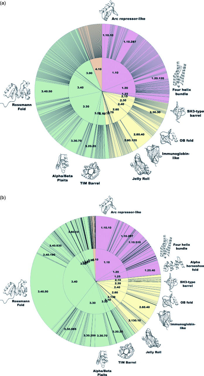
The CATH database of protein domain structures (http://www.biochem.ucl.ac.uk/bsm/cath/) currently contains 43,229 domains classified into 1467 superfamilies and 5107 sequence families. Each structural family is expanded with sequence relatives from GenBank and completed genomes, using a variety of efficient sequence search protocols and reliable thresholds. This extended CATH protein family database contains 616,470 domain sequences classified into 23,876 sequence families. This results in the significant expansion of the CATH HMM model library to include models built from the CATH sequence relatives, giving a 10% increase in coverage for detecting remote homologues. An improved Dictionary of Homologous superfamilies (DHS) (http://www.biochem.ucl.ac.uk/bsm/dhs/) containing specific sequence, structural and functional information for each superfamily in CATH considerably assists manual validation of homologues. Information on sequence relatives in CATH superfamilies, GenBank and completed genomes is presented in the CATH associated DHS and Gene3D resources. Domain partnership information can be obtained from Gene3D (http://www.biochem.ucl.ac.uk/bsm/cath/Gene3D/). A new CATH server has been implemented (http://www.biochem.ucl.ac.uk/cgi-bin/cath/CathServer.pl) providing automatic classification of newly determined sequences and structures using a suite of rapid sequence and structure comparison methods. The statistical significance of matches is assessed and links are provided to the putative superfamily or fold group to which the query sequence or structure is assigned.
Figures
References
-
- Bray J.E., Todd,A.E., Pearl,F.M., Thornton,J.M. and Orengo,C.A. (2000) The CATH Dictionary of Homologous Superfamilies (DHS): a consensus approach for identifying distant structural homologues. Protein Eng., 13, 153–165. - PubMed
-
- Taylor W. and Orengo,C. (1989) Protein structure alignment. J. Mol. Biol., 208, 1–22. - PubMed
Publication types
MeSH terms
Substances
LinkOut - more resources
Full Text Sources
Other Literature Sources
