. 2005 Jan;17(1):311-25.
doi: 10.1105/tpc.104.027235. Epub 2004 Dec 17.

A NAC domain protein interacts with tomato leaf curl virus replication accessory protein and enhances viral replication

Affiliations

A NAC domain protein interacts with tomato leaf curl virus replication accessory protein and enhances viral replication

Luke A Selth et al. Plant Cell. 2005 Jan.

Abstract

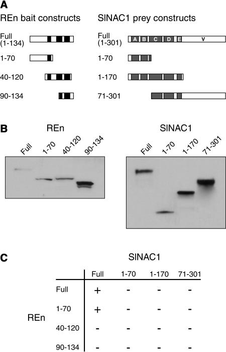
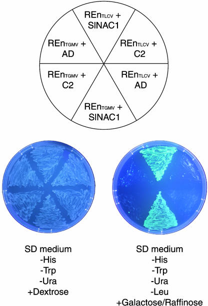
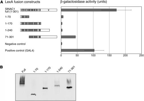
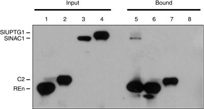
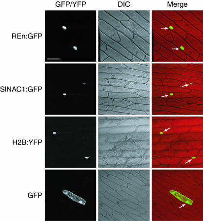
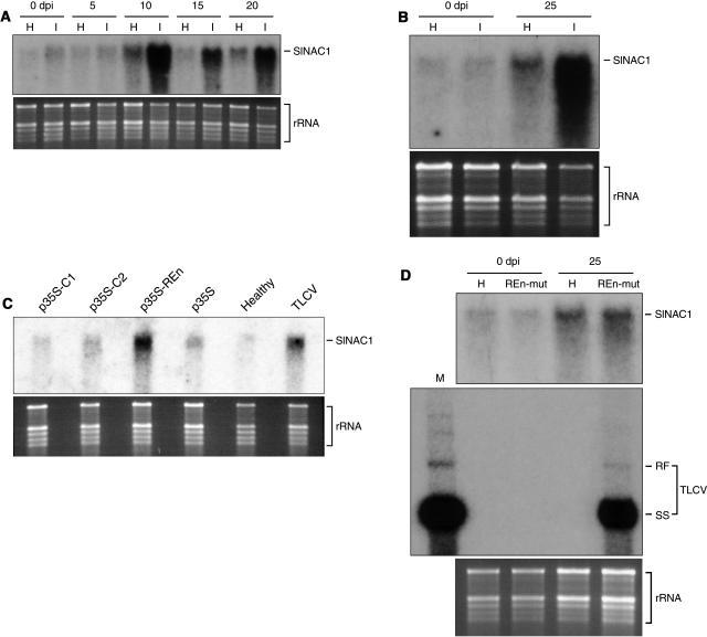
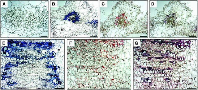
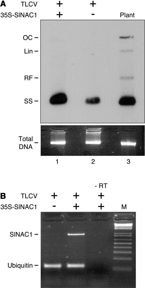
Geminivirus replication enhancer (REn) proteins dramatically increase the accumulation of viral DNA species by an unknown mechanism. In this study, we present evidence implicating SlNAC1, a new member of the NAC domain protein family from tomato (Solanum lycopersicum), in Tomato leaf curl virus (TLCV) REn function. We isolated SlNAC1 using yeast (Saccharomyces cerevisiae) two-hybrid technology and TLCV REn as bait, and confirmed the interaction between these proteins in vitro. TLCV induces SlNAC1 expression specifically in infected cells, and this upregulation requires REn. In a transient TLCV replication system, overexpression of SlNAC1 resulted in a substantial increase in viral DNA accumulation. SlNAC1 colocalized with REn to the nucleus and activated transcription of a reporter gene in yeast, suggesting that in healthy cells it functions as a transcription factor. Together, these results imply that SlNAC1 plays an important role in the process by which REn enhances TLCV replication.


Figures

Figure 1.
Figure 1.
Nucleotide Sequence of SlNAC1 and Alignment of Its Putative Translation Product with Other NAC Domain Proteins. (A) Nucleotide and amino acid sequences of SlNAC1. The five subdomains (A to E) comprising the NAC domain are shown in empty boxes. A putative nuclear localization signal is indicated by a bold line under the sequence PRDRKYP. The TAR-CM of the ATAF subgroup is also boxed. (B) The predicted amino acid sequence of SlNAC1 (Figure 1A) and known NAC family proteins were subjected to phylogenetic analysis. Multiple sequence alignment of the proteins was conducted using ClustalX (Thompson et al., 1997), and phylogenetic analysis was performed by the neighbor-joining method (Saitou and Nei, 1987). A bootstrap analysis of 1000 resampling replicates was conducted with ClustalX. The rooted phylogenetic tree was displayed using the NJPlot program included with ClustalX. The gene names and references for other NACs are as follows: A. thaliana, ATAF1 and ATAF2 (Aida et al., 1997), AtNAC2 (Takada et al., 2001), AtNAC3 (Takada et al., 2001), AtNAM (Duval et al., 2002), CUC1 (Takada et al., 2001), CUC2 (Takada et al., 2001), CUC3 (Vroemen et al., 2003), NAC1 (Xie et al., 2000), NAC2, NAP (Sablowski and Meyerowitz, 1998), and TIP (Ren et al., 2000); rice, OsNAC1 to OsNAC8 (Kikuchi et al., 2000); petunia, NAM (Souer et al., 1996); tomato, SenU5 (John et al., 1997); potato, StNAC (Collinge and Boller, 2001); and wheat, GRAB1 and GRAB2 (Xie et al., 1999).
Figure 2.
Figure 2.
Deletion Analysis of REn and SlNAC1 to Identify Regions Required for Interaction between the Two Proteins. (A) Diagrammatic representation of REn (bait) and SlNAC1 proteins (prey) tested for interaction. The REn proteins were expressed as LexA DNA BD fusions, and the SlNAC1 proteins were expressed as B42 AD fusions. The positions of three putative α-helices in REn are indicated by closed boxes. In SlNAC1, the positions of the NAC subdomains are shown in shaded boxes (A to E), whereas the variable C terminus is denoted V. (B) Immunoblot analysis of yeast cells demonstrating that noninteracting REn-LexA fusions and SlNAC1-B42 fusions are expressed at levels similar to those of interacting fusion proteins. Total protein from yeast cultures containing different REn and SlNAC1 fusion proteins was extracted, fractionated on 4 to 20% SDS-polyacrylamide gels, and immunoblotted with anti-LexA (to detect REn-LexA fusions) or anti-hemagglutinin (HA) (to detect SlNAC1-B42 fusions). (C) The N-terminal region of REn is important for SlNAC1 binding. Interaction was indicated by the ability of cells transformed with bait, prey, and displayREPORTER plasmids to grow on medium lacking Leu. As an additional indicator of interaction, colonies were monitored for GFP expression by visualization under UV light.
Figure 3.
Figure 3.
SlNAC1 Interacts with Both TLCV and TGMV REn. Yeast two-hybrid assays testing the ability of SlNAC1 to interact with REn of TLCV (REnTLCV) and TGMV (REnTGMV). Yeast coexpressing proteins as indicated (top) were grown on SD – His – Trp – uracil (Ura) medium (bottom left), and interaction was tested by Leu prototrophy and GFP expression on an inductive carbon source (galactose and raffinose; bottom right). REn proteins were fused to the LexA DNA BD, whereas SlNAC1 was fused to the B42 AD. Negative controls included REnTLCV and REnTGMV coexpressed with TLCV C2 fused to the AD, or coexpressed with AD alone.
Figure 4.
Figure 4.
The Divergent C-Terminal Region of SlNAC1 Is Able to Activate Transcription in Yeast. (A) Regions of SlNAC1 able to activate transcription in yeast. The LexA:SlNAC1 fusion proteins are represented diagrammatically at left, with the positions of the NAC subdomains shown in shaded boxes (A to E) and the variable C terminus denoted V. The ability of LexA:SlNAC1 fusion proteins to activate transcription in yeast is shown at right. Activities were assayed by measuring β-galactosidase activity in total protein extracts from cells containing pLexA-SlNAC1 plasmids and pSH18-34, which contains a lacZ reporter gene downstream of the LexA recognition site. Positive control corresponds to yeast containing pSH18-34 and expressing a LexA fusion with the GAL4 AD. Negative control corresponds to yeast containing pSH18-34 and expressing LexA. Error bars indicate the standard deviation for each sample. (B) Immunoblot analysis of yeast cells demonstrating that nontransactivating LexA:SlNAC1 fusions are expressed at levels similar to those of transactivating fusion proteins. Total protein from yeast cultures containing fusion proteins was extracted, fractionated on SDS-polyacrylamide gels, and immunoblotted with anti-LexA.
Figure 5.
Figure 5.
REn Interacts with SlNAC1 in Vitro. Purified 6×His-tagged proteins were mixed with crude CBP-tagged protein mixtures, incubated with Ni-NTA, and washed extensively to remove any unbound protein. Bound protein was resuspended in loading buffer, resolved by SDS-PAGE, and analyzed by immunoblotting using anti-polyHis and anti-FLAG (CBP-tagged proteins also contain a FLAG epitope) antibodies. Reactions were as follows: 6×His-REn and CBP-SlNAC1 (lane 5), 6×His-REn and CBP-SlUPTG1 (lane 6), 6×His-C2 and CBP-SlNAC1 (lane 7), and CBP-SlNAC1 alone (lane 8). Protein inputs for each reaction are shown: 6×His-REn (lane 1), 6×His-C2 (lane 2), CBP-SlNAC1 (lane 3), and CBP-SlUPTG1 (lane 4).
Figure 6.
Figure 6.
REn and SlNAC1 Localize to the Nucleus of Onion Cells. REn:GFP (top row) and SlNAC1:GFP (second row), as well as GFP alone (bottom row), were expressed in onion epidermal cells using the CaMV 35S promoter after biolistic delivery of vector DNA. A positive control for nuclear localization, H2B:YFP, is also shown (third row). Cells were analyzed for GFP and YFP fluorescence (left column) by confocal microscopy. Differential interference contrast (DIC) images and merge images are shown in the middle and right columns, respectively. Nuclei in merge images are indicated by arrows. Bar = 100 μm.
Figure 7.
Figure 7.
SlNAC1 Is Induced by TLCV Infection. (A) TLCV infection results in an upregulation of SlNAC1 gene expression. RNA gel blot showing the expression of SlNAC1 in healthy (H) or TLCV-infected (I) tomato plants. Tissue samples were obtained at 0, 5, 10, 15, and 20 dpi. (B) TYLCSV infection results in an upregulation of SlNAC1 gene expression. RNA gel blot showing the expression of SlNAC1 in healthy or TYLCSV-infected tomato plants. Tissue samples were obtained 0 and 25 dpi. (C) Transient expression of REn is sufficient to induce SlNAC1 gene expression. Tomato leaves were infiltrated with A. tumefaciens cells containing a replication-competent TLCV 1.1mer, p35S, or p35S expressing the TLCV genes C1, C2, and REn. RNA was extracted from tissues 5 d postinfiltration and SlNAC1 expression analyzed by RNA gel blotting. (D) A TLCV REn mutant cannot induce SlNAC1 gene expression. RNA gel blot showing the expression of SlNAC1 in healthy plants or plants infected with a TLCV REn mutant (REn-mut) at 0 and 25 dpi (top). The presence of replicating TLCV REn mutant was confirmed by DNA gel blotting (middle). In this blot, we also ran an extract obtained from plants infected with wild-type virus (left, designated M); the ratio of REn mutant:wild-type virus total nucleic acid extracts is 20:1. TLCV DNA species are marked RF (supercoiled double-stranded replicative form) and SS (single stranded).
Figure 8.
Figure 8.
Induction of SlNAC1 by TLCV Occurs Only in Infected Cells. Tissue sections derived from mock-inoculated (A) and TLCV-infected ([B] to [G]) leaves of N. benthamiana were hybridized with either fluorescein-labeled ssRNA probe complementary to SlNAC1 ([A], [C], [D], [F], and [G]) or DIG-labeled ssRNA probe complementary to TLCV ([A], [B], [D], [E], and [G]). (A) to (D) are cross sections and (E) to (G) are longitudinal sections taken from the main leaf vein. Bar = 100 μm. Cell types present are indicated: E, epidermal; M, mesophyll; P, phloem; and X, xylem.
Figure 9.
Figure 9.
SlNAC1 Expression Enhances TLCV ssDNA Accumulation. (A) Expression of SlNAC1 enhances TLCV ssDNA accumulation in a transient replication assay. A. tumefaciens cells harboring Bin19-TLCV1.1 were combined with A. tumefaciens cells containing either an empty expression construct (lane 2) or p35S-SlNAC1 (lane 1) and cocultivated for 48 h with leaf strips from N. benthamiana plants. DNA was extracted from tissue samples 3 d later and replication of TLCV analyzed by DNA gel blotting. Lane 3 (Plant) is a sample extracted from TLCV-infected N. benthamiana used as a marker for TLCV DNA forms, marked OC (open circular double stranded), Lin (linear double stranded), RF (supercoiled double-stranded replicative form), and SS (single stranded). OC, Lin and RF DNA forms were observed in extracts from N. benthamiana leaf strips after longer exposures. (B) Analysis of SlNAC1 expression by p35S-SlNAC1 in N. benthamiana leaf strips by semiquantitative RT-PCR. Total RNA was prepared from leaf strips treated with TLCV plus an empty expression construct or TLCV plus p35S-SlNAC1. Ubiquitin mRNA served as an internal control. RT reaction mix without reverse transcriptase was used as a negative control (marked –RT). M, size markers.

References

    1. Abouzid, A.M., Frischmuth, T., and Jeske, H. (1988). A putative replicative form of the Abutilon mosaic virus (gemini group) in a chromatin-like structure. Mol. Gen. Genet. 212, 252–258.
    1. Ach, R.A., Durfee, T., Miller, A.B., Taranto, P., Hanley-Bowdoin, L., Zambryski, P.C., and Gruissem, W. (1997). RRB1 and RRB2 encode maize retinoblastoma-related proteins that interact with a plant D-type cyclin and geminivirus replication protein. Mol. Cell. Biol. 17, 5077–5086. - PMC - PubMed
    1. Aida, M., Ishida, T., Fukaki, H., Fujisawa, H., and Tasaka, M. (1997). Genes involved in organ separation in Arabidopsis: An analysis of the cup-shaped cotyledon mutant. Plant Cell 9, 841–857. - PMC - PubMed
    1. Behjatnia, S.A.A., Dry, I.B., and Rezaian, M.A. (1998). Identification of the replication-associated protein binding domain within the intergenic region of tomato leaf curl geminivirus. Nucleic Acids Res. 26, 925–931. - PMC - PubMed
    1. Boisnard-Lorig, C., Colon-Carmona, A., Bauch, M., Hodge, S., Doerner, P., Bancharel, E., Dumas, C., Haseloff, J., and Berger, F. (2001). Dynamic analyses of the expression of the HISTONE∷YFP fusion protein in arabidopsis show that syncytial endosperm is divided in mitotic domains. Plant Cell 13, 495–509. - PMC - PubMed

Publication types

MeSH terms

Associated data

LinkOut - more resources