. 2005 Jan;6(1):39-45.
doi: 10.1038/sj.embor.7400314.

How the human telomeric proteins TRF1 and TRF2 recognize telomeric DNA: a view from high-resolution crystal structures

Affiliations

How the human telomeric proteins TRF1 and TRF2 recognize telomeric DNA: a view from high-resolution crystal structures

Robert Court et al. EMBO Rep. 2005 Jan.

Erratum in

  • EMBO Rep. 2005 Feb;6(2):191

Abstract

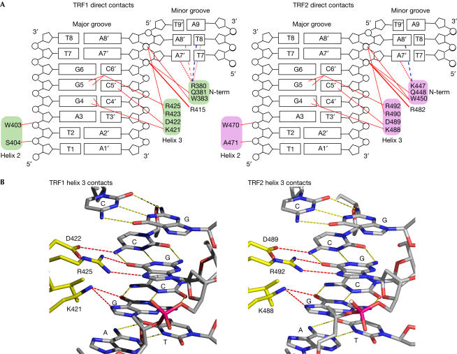
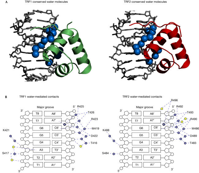
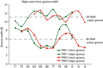
Human telomeres consist of tandem arrays of TTAGGG sequence repeats that are specifically bound by two proteins, TRF1 and TRF2. They bind to DNA as preformed homodimers and have the same architecture in which the DNA-binding domains (Dbds) form independent structural units. Despite these similarities, TRF1 and TRF2 have different functions at telomeres. The X-ray crystal structures of both TRF1- and TRF2-Dbds in complex with telomeric DNA (2.0 and 1.8 angstroms resolution, respectively) show that they recognize the same TAGGGTT binding site by means of homeodomains, as does the yeast telomeric protein Rap1p. Two of the three G-C base pairs that characterize telomeric repeats are recognized specifically and an unusually large number of water molecules mediate protein-DNA interactions. The binding of the TRF2-Dbd to the DNA double helix shows no distortions that would account for the promotion of t-loops in which TRF2 has been implicated.


Figures

Figure 1
Figure 1
Protein and DNA constructs used in the crystallization of the TRF1-Dbd and TRF2-Dbd complexes. (A) Schematic representation of the TRF1 and TRF2 proteins indicating the location of the two domains of sequence homology: the TRFH dimerization domain (yellow) and the Myb DNA-binding motif (red). The percentage sequence identity and homology are shown. (B) TRF1 and TRF2 protein constructs used in crystallization. The sequences of the two Dbds have been aligned, and identical (marked with an asterisk) and similar (marked with a dot) residues between TRF1 and TRF2 are denoted. The open rectangles indicate the position of the α-helices. In each case, the N-terminus of the protein is disordered, and the first residue for which there is electron density is residue R380 in TRF1 and K447 in TRF2. (C) The two DNA binding sites used in the crystals. The two sets of three G-C base pairs present in each binding site are highlighted in magenta.
Figure 2
Figure 2
Global structure of two TRF2-Dbds bound to double-stranded telomeric DNA. The TRF1-Dbd–DNA complex has the same overall structure and hence is not shown. The protein molecules in the complex are shown as a ribbon representation. The two sets of three G-C base pairs present in the DNA binding site are highlighted in magenta. All figures of the structures were created using Pymol (DeLano, 2002).
Figure 3
Figure 3
Summary of direct protein–DNA contacts in the TRF1-Dbd–DNA and TRF2-Dbd–DNA complexes. (A) Maps of protein–DNA contacts. The DNA is represented as an opened-out helix. Red lines indicate direct hydrogen bonds. These contacts are conserved between the two molecules in one complex. Direct contacts in the minor groove made by residues R380 of TRF1 and K447 of TRF2 that differ in the two protein molecules in one complex are indicated by dotted and dashed red lines. A dashed blue line depicts water-mediated contacts. (B) Views at atomic resolution of the hydrogen bonds between residues in the DNA-recognition helix and the bases in the major groove of DNA.
Figure 4
Figure 4
Summary of water-mediated protein–DNA contacts in the TRF1-Dbd–DNA and TRF2-Dbd–DNA complexes. (A) Conservation in the water structure at the protein–DNA interface. The two proteins are shown as ribbon representations and only half of each complex is shown. Conserved water molecules at the protein–DNA interface are represented by blue spheres. (B) Summary of water-mediated contacts in TRF1-Dbd–DNA and TRF2-Dbd–DNA complexes. Conserved water molecules are shown in blue and additional water molecules are shown in yellow. Blue dashed lines depict the network of water-mediated hydrogen bonds.
Figure 5
Figure 5
DNA structure in the TRF1 and TRF2 binding sites. The widths of the DNA major and minor grooves in the complexes were calculated using the program CURVES (Lavery & Sklenar, 1989). The average major and minor groove widths of B-form DNA are shown for reference.

References

    1. Abrahams JP, Leslie AGW (1996) Methods used in the structure determination of bovine mitochondrial F-1 ATPase. Acta Crystallogr D 52: 30–42 - PubMed
    1. Ancelin K, Brun C, Gilson E (1998) Role of the telomeric DNA-binding protein TRF2 in the stability of human chromosome ends. BioEssays 20: 879–883 - PubMed
    1. Bianchi A, Smith S, Chong L, Elias P, de Lange T (1997) TRF1 is a dimer and bends telomeric DNA. EMBO J 16: 1785–1794 - PMC - PubMed
    1. Bianchi A, Stansel RM, Fairall L, Griffith JD, Rhodes D, de Lange T (1999) TRF1 binds a bipartite telomeric site with extreme spatial flexibility. EMBO J 18: 5735–5744 - PMC - PubMed
    1. Bilaud T, Koering CE, Binet-Brasselet E, Ancelin K, Pollice A, Gasser SM, Gilson E (1996) The telobox, a Myb-related telomeric DNA binding motif found in proteins from yeast, plants and human. Nucleic Acids Res 24: 1294–1303 - PMC - PubMed

MeSH terms