. 2004 Dec 28;43(51):16161-73.
doi: 10.1021/bi049017a.

GRP1 pleckstrin homology domain: activation parameters and novel search mechanism for rare target lipid

Affiliations

GRP1 pleckstrin homology domain: activation parameters and novel search mechanism for rare target lipid

John A Corbin et al. Biochemistry. .

Abstract

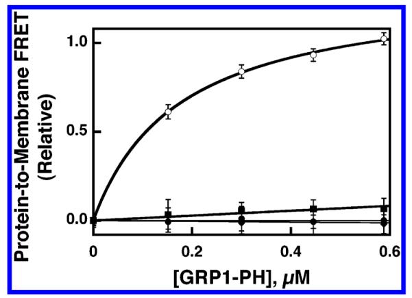
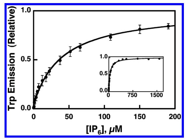
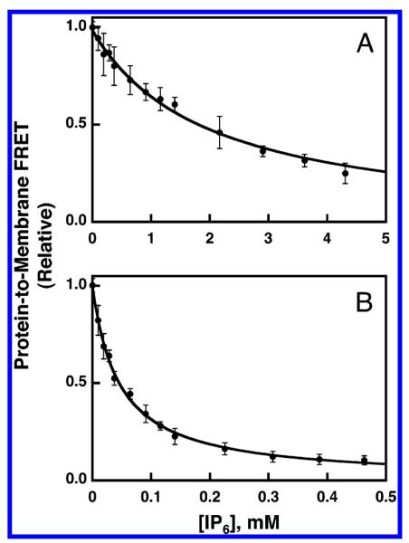
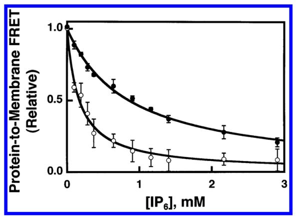
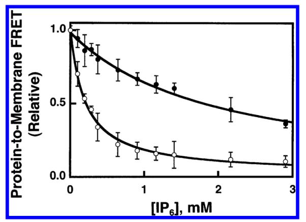
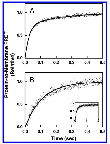
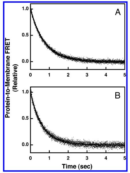
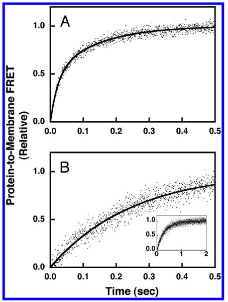
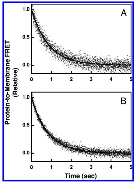
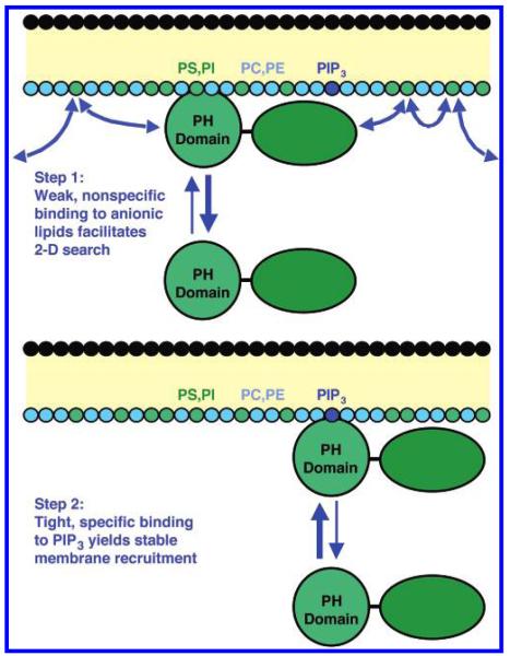
Pleckstrin homology (PH) domains play a central role in a wide array of signaling pathways by binding second messenger lipids of the phosphatidylinositol phosphate (PIP) lipid family. A given type of PIP lipid is formed in a specific cellular membrane where it is generally a minor component of the bulk lipid mixture. For example, the signaling lipid PI(3,4,5)P(3) (or PIP(3)) is generated primarily in the inner leaflet of the plasma membrane where it is believed to never exceed 0.02% of the bulk lipid. The present study focuses on the PH domain of the general receptor for phosphoinositides, isoform 1 (GRP1), which regulates the actin cytoskeleton in response to PIP(3) signals at the plasma membrane surface. The study systematically analyzes both the equilibrium and kinetic features of GRP1-PH domain binding to its PIP lipid target on a bilayer surface. Equilibrium binding measurements utilizing protein-to-membrane fluorescence resonance energy transfer (FRET) to detect GRP1-PH domain docking to membrane-bound PIP lipids confirm specific binding to PIP(3). A novel FRET competitive binding measurement developed to quantitate docking affinity yields a K(D) of 50 +/- 10 nM for GRP1-PH domain binding to membrane-bound PIP(3) in a physiological lipid mixture approximating the composition of the plasma membrane inner leaflet. This observed K(D) lies in a suitable range for regulation by physiological PIP(3) signals. Interestingly, the affinity of the interaction decreases at least 12-fold when the background anionic lipids phosphatidylserine (PS) and phosphatidylinositol (PI) are removed from the lipid mixture. Stopped-flow kinetic studies using protein-to-membrane FRET to monitor association and dissociation time courses reveal that this affinity decrease arises from a corresponding decrease in the on-rate for GRP1-PH domain docking with little or no change in the off-rate for domain dissociation from membrane-bound PIP(3). Overall, these findings indicate that the PH domain interacts not only with its target lipid, but also with other features of the membrane surface. The results are consistent with a previously undescribed type of two-step search mechanism for lipid binding domains in which weak, nonspecific electrostatic interactions between the PH domain and background anionic lipids facilitate searching of the membrane surface for PIP(3) headgroups, thereby speeding the high-affinity, specific docking of the domain to its rare target lipid.


Figures

Figure 1
Figure 1
Equilibrium binding of GRP1-PH domain to synthetic lipid vesicles containing different PIP lipids. Sonicated lipid vesicles possessing the indicated PIP lipid exposed on their surfaces were titrated with isolated GRP1-PH domain (1 μM) at 25 °C. The docking of GRP1-PH domain to the vesicles was detected by protein-to-membrane FRET using intrinsic protein tryptophan residues as donors and membrane-bound dansyl-phosphatidylethanolamine lipids as acceptors (see Experimental Procedures). Vesicles were composed of PE/PC/PS/PI/dPE/SM/PIPx (41.4/18.4/18.4/9.2/5/4.6/3 mol %) where the total accessible lipid and accessible PIP lipid concentrations were 59 and 1.8 μM, respectively, or PE/PC/PS/PI/dPE/SM (42.75/19/19/9.5/5/4.7/5 mol %) where the total accessible lipid was 59 μM, and the final buffer composition was 140 mM KCl, 15 mM NaCl, 1 mM MgCl2, 10 mM DTT, 25 mM HEPES, pH 7.4. Individual samples contained PI(3,4,5)P3 (○), PI(3,4)P2 (■), PI(4,5)P2 (▲), or no PIP lipid (●). Error bars represent the standard deviation of the mean for three replicate experiments.
Figure 2
Figure 2
Equilibrium binding of IP6 to isolated GRP1 PH domain in the absence of membranes. Free GRP1-PH domain (0.8 μM) was titrated at 25 °C with increasing concentrations of IP6. Binding of IP6 to the domain was monitored via the observed increase in intrinsic tryptophan fluorescence (see Experimental Procedures). The final buffer composition was 140 mM KCl, 15 mM NaCl, 1 mM MgCl2, 1 mM DTT, 25 mM HEPES, pH 7.4). Error bars represent the standard deviation of the mean for four replicate experiments. The solid curve represents the nonlinear least-squares best fit for a homogeneous population of independent IP6 binding sites (eq 1). Inset shows the same data over the full IP6 concentration range used to calculate the binding constant.
Figure 3
Figure 3
Use of FRET competitive binding assay to measure the equilibrium affinity of GRP1-PH domain for membrane-bound PIP lipids. A preformed complex between GRP1-PH domain (0.75 μM) and sonicated lipid vesicles containing the indicated PIP lipid (101 μM total accessible lipid; 3 μM accessible PIP lipid) was titrated at 25 °C with increasing concentrations of IP6. The competitive displacement of GRP1-PH domain from the membrane surface was monitored as decreasing protein-to-membrane FRET (see Experimental Procedures). The final buffer composition was 140 mM KCl, 15 mM NaCl, 1 mM MgCl2, 10 mM DTT, and 25 mM HEPES, pH 7.4. Membranes contained (A) PE/PC/PS/PI/dPE/SM/PI(3,4,5)-P3 (41.4/18.4/18.4/9.2/5/4.6/3 mol %) or (B) PE/PC/PS/PI/dPE/SM/PI(3,4)P2 membranes (41.4/18.4/18.4/9.2/5/4.6/3 mol %). Error bars represent the standard deviation of the mean for at least three replicate experiments. Solid curves represent nonlinear least-squares best fits for a homogeneous population of binding sites (eq 2), which yield the apparent IP6 affinity, enabling calculation of the KD for PH domain docking to membrane-bound PIP lipid (eq 6, Table 2).
Figure 4
Figure 4
Use of FRET competitive binding assay to measure the equilibrium affinity of GRP1-PH domain for membrane-bound PIP3 in a simple lipid mixture. A preformed complex between GRP1-PH domain (0.75 μM) and sonicated lipid vesicles containing PI-(3,4,5)P3 (101 μM total accessible lipid; 3 μM accessible PI(3,4,5)-P3) was titrated at 25 °C with increasing concentrations of IP6. The competitive displacement of GRP1-PH domain from the membrane surface was monitored as decreasing protein-to-membrane FRET (see Experimental Procedures). The final buffer composition was 140 mM KCl, 15 mM NaCl, 1 mM MgCl2, 10 mM DTT, 25 mM HEPES, pH 7.4, and membranes contained one of the two following lipid mixtures: PC/PS/dPE/PI(3,4,5)P3 (69/23/5/3 mol %) (●) or PC/dPE/PI(3,4,5)P3 (92/5/3 mol %) (○). Error bars represent the standard deviation of the mean for at least three replicate experiments. Solid curves represent nonlinear least-squares best fits for a homogeneous population of binding sites (eq 2), which yield the apparent IP6 affinity, enabling calculation of the KD for PH domain docking to membrane-bound PI(3,4,5)P3 lipid (eq 6, Table 2).
Figure 5
Figure 5
Use of FRET competitive binding assay to measure the equilibrium affinity of GRP1-PH domain for membrane-bound PIP3 in a physiological lipid mixture. A preformed complex between GRP1-PH domain (0.75 μM) and sonicated lipid vesicles containing PI(3,4,5)P3 (101 μM total accessible lipid; 3 μM accessible PI(3,4,5)P3) was titrated at 25 °C with increasing concentrations of IP6. The competitive displacement of GRP1-PH domain from the membrane surface was monitored as decreasing protein-to-membrane FRET (see Experimental Procedures). The final buffer composition was 140 mM KCl, 15 mM NaCl, 1 mM MgCl2, 10 mM DTT, 25 mM HEPES, pH 7.4, and membranes contained one of the two following lipid mixtures: PE/PC/PS/PI/dPE/SM/PI-(3,4,5)P3 (41.4/18.4/18.4/9.2/5/4.6/3 mol %) (●) or PE/PC/SM/dPE/PI(3,4,5)P3 (59.1/26.3/6.6/5/3 mol %) (○). Error bars represent the standard deviation of the mean for at least three replicate experiments. Solid curves represent nonlinear least-squares best fits for a homogeneous population of binding sites (eq 2), which yield the apparent IP6 affinity, enabling calculation of the KD for PH domain docking to membrane-bound PI(3,4,5)P3 lipid (eq 6, Table 2).
Figure 6
Figure 6
Association kinetics of GRP1-PH domain docking to membrane-bound PIP3 in a simple lipid mixture. The association reaction was triggered by rapid mixing of GRP1-PH domain with sonicated lipid vesicles containing PI(3,4,5)P3 at 25 °C in a stopped-flow spectrofluorimeter while monitoring the increasing protein-to-membrane FRET (see Experimental Procedures). The GRP1-PH domain and lipid concentrations following mixing were equivalent to those used in the equilibrium experiments (0.75 μM GRP1-PH domain; 101 μM total lipid accessible; 3 μM PI(3,4,5)P3 accessible). The buffer contained 140 mM KCl, 15 mM NaCl, 1 mM MgCl2, 1 mM DTT, and 25 mM HEPES, pH 7.4, and the membranes were composed of (A) PC/PS/dPE/PI(3,4,5)P3 (69/23/5/3 mol %) or (B) PC/dPE/PI(3,4,5)P3 (92/5/3 mol %). Data points represent the average values from at least six separate replicate timecourses. The solid curves represent the best fit to (A) a double-exponential equation or (B) a single-exponential equation (eq 3 or 4, respectively). Panel B inset shows the same data over the full time course used to determine kon.
Figure 7
Figure 7
Dissociation kinetics of GRP1-PH domain docked to membrane-bound PIP3 in a simple lipid mixture. The dissociation reaction was triggered by rapid mixing of IP6 with GRP1-PH domain bound to PI(3,4,5)P3 on vesicles in a stopped-flow fluorimeter at 25 °C. The resulting dissociation of GRP1-PH domain from the vesicles was monitored by protein-to-membrane FRET (see Experimental Procedures). The GRP1-PH domain and lipid concentrations following mixing were equivalent to those used in the equilibrium experiments (0.75 μM GRP1-PH; 101 μM total lipid accessible; 3 μM PI(3,4,5)P3 accessible). The buffer contained 140 mM KCl, 15 mM NaCl, 1 mM MgCl2, 10 mM DTT, and 25 mM HEPES, pH 7.4, and the membranes were composed of (A) PC/PS/dPE/PI(3,4,5)P3 (69/23/5/3 mol %) or (B) PC/dPE/PI(3,4,5)P3 (92/5/3 mol %). Data points represent the average values from at least six separate replicate time courses. The solid curves represent the best fits to a single-exponential equation (eq 5).
Figure 8
Figure 8
Association kinetics of GRP1-PH domain docking to membrane-bound PIP3 in a physiological lipid mixture. The association reaction was triggered by rapid mixing of GRP1-PH domain with sonicated lipid vesicles containing PI(3,4,5)P3 at 25 °C in a stopped-flow spectrofluorimeter while monitoring the increasing protein-to-membrane FRET (see Experimental Procedures). The GRP1-PH domain and lipid concentrations following mixing were equivalent to those used in the equilibrium experiments (0.75 μM GRP1-PH domain: 101 μM total lipid accessible; 3 μM PI(3,4,5)P3 accessible). The buffer contained 140 mM KCl, 15 mM NaCl, 1 mM MgCl2, 10 mM DTT, and 25 mM HEPES, pH 7.4, and the membranes were composed of (A) PE/PC/PS/PI/dPE/SM/PI(3,4,5)P3 (41.4/18.4/18.4/9.2/5/4.6/3 mol %) or (B) PE/PC/SM/dPE/PI(3,4,5)P3 (59.1/26.3/6.6/5/3 mol %). Data points represent the average values from at least six separate replicate time courses. The solid curves represent the best fit to (A) a double-exponential equation or (B) a single-exponential equation (eq 3 or 4, respectively). Panel B inset shows the same data over the full time course used to determine kon.
Figure 9
Figure 9
Dissociation kinetics of GRP1-PH domain docked to membrane-bound PIP3 in a physiological lipid mixture. The dissociation reaction was triggered by rapid mixing of IP6 with GRP1-PH domain bound to PI(3,4,5)P3 on vesicles in a stopped-flow fluorimeter at 25 °C. The resulting dissociation of GRP1-PH domain from the vesicles was monitored by protein-to-membrane FRET (see Experimental Procedures). The GRP1-PH domain and lipid concentrations following mixing were equivalent to those used in the equilibrium experiments (0.75 μM GRP1-PH domain; 101 μM total lipid accessible; 3 μM PI(3,4,5)P3 accessible). The buffer contained 140 mM KCl, 15 mM NaCl, 1 mM MgCl2, 10 mM DTT, and 25 mM HEPES, pH 7.4, and the membranes were composed of (A) PE/PC/PS/PI/dPE/SM/PI(3,4,5)P3 (41.4/18.4/18.4/9.2/5/4.6/3 mol %) or (B) PE/PC/SM/dPE/PI(3,4,5)P3 (59.1/26.3/6.6/5/3 mol %). Data points represent the average values from at least six separate replicate time courses. The solid curves represent the best fits to a single-exponential equation (eq 5).
Figure 10
Figure 10
Model of the search mechanism used by GRP1-PH domain to find the rare target lipid PIP3. Shown is a schematic plasma membrane consisting of a core hydrocarbon region (yellow), the headgroups of the outer leaflet (black) and the headgroups of the cytoplasmic inner leaflet color coded according to chemical identity (cyan for neutral headgroups, green for the anionic lipids PS and PI, and blue for PI(3,4,5)P3). Also shown is the full-length GRP1 protein with its distinct PH and GEF (guanine-nucleotide exchange factor) domains. Panel A depicts step 1: The PH domain of cytosolic GRP1 forms transient weak electrostatic interactions with the abundant background anionic lipids PS and PI. The nonspecific electrostatic interactions with PS and PI are not sufficient to generate stable docking of GRP1 to the membrane but are sufficient to significantly increase the residency time of the protein at or near the membrane. This increased residency time near the membrane facilitates electrostatic steering, as well as a short-range two-dimensional search across the membrane surface for the rare target PIP3. Yet the membrane association is transient, and the bold arrow emphasizes that the equilibrium favors the free cytosolic state of the protein, while the curved arrows indicate that the short intervals of the two-dimensional search are frequently interrupted by dissociation events. Panel B depicts step 2: Once its target lipid has been found, the PH domain of GRP1 binds specifically and with high affinity to PIP3 resulting in stable membrane docking. The bold arrow emphasizes that the equilibrium favors the membrane-bound state of the protein. The dissociation event is dominated by the interaction with PIP3 and perhaps additional hydrophobic interactions with the surrounding membrane, but electrostatic interactions with the surrounding membrane appear to be minor by comparison since background anionic lipids have little effect on the dissociation kinetics (see text).

Similar articles

Cited by

References

    1. Yu JW, Mendrola JM, Audhya A, Singh S, Keleti D, DeWald DB, Murray D, Emr SD, Lemmon MA. Genome-wide analysis of membrane targeting by S. cerevisiae pleckstrin homology domains. Mol. Cell. 2004;13:677–88. - PubMed
    1. Czech MP. Dynamics of phosphoinositides in membrane retrieval and insertion. Annu. ReV. Physiol. 2003;65:791–815. - PubMed
    1. Luo J, Manning BD, Cantley LC. Targeting the PI3K-Akt pathway in human cancer: rationale and promise. Cancer Cell. 2003;4:257–62. - PubMed
    1. Hurley JH, Meyer T. Subcellular targeting by membrane lipids. Curr. Opin. Cell Biol. 2001;13:146–52. - PubMed
    1. Vanhaesebroeck B, Leevers SJ, Ahmadi K, Timms J, Katso R, Driscoll PC, Woscholski R, Parker PJ, Waterfield MD. Synthesis and function of 3-phosphorylated inositol lipids. Annu. ReV. Biochem. 2001;70:535–602. - PubMed

Publication types

MeSH terms