. 2005 May 1;387(Pt 3):897-903.
doi: 10.1042/BJ20041312.

Misfolded BiP is degraded by a proteasome-independent endoplasmic-reticulum-associated degradation pathway

Affiliations

Misfolded BiP is degraded by a proteasome-independent endoplasmic-reticulum-associated degradation pathway

Gerda Donoso et al. Biochem J. .

Abstract

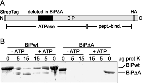
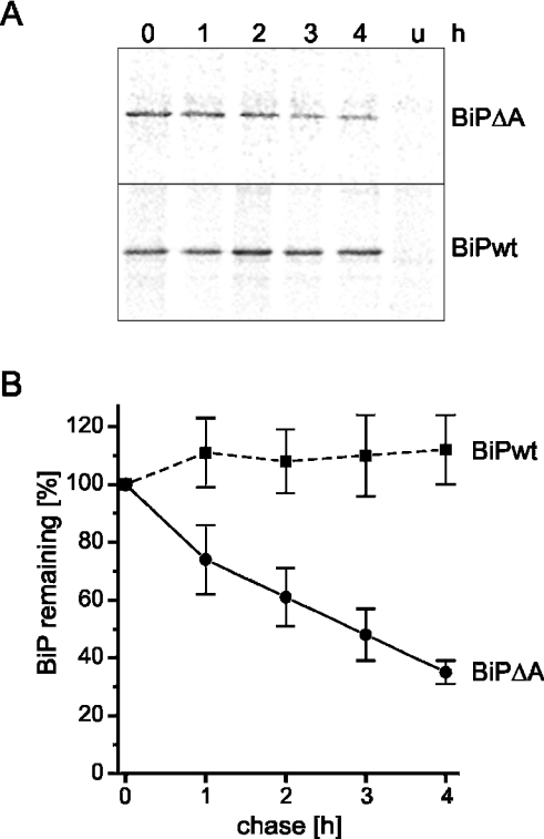
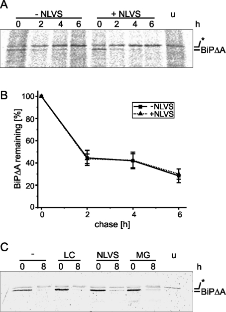
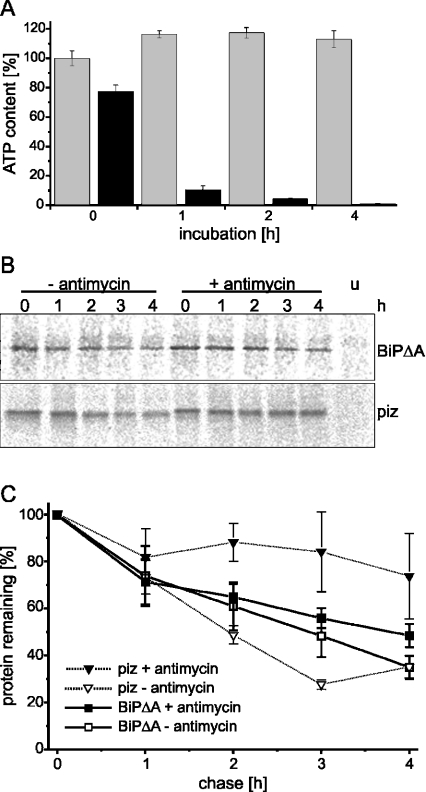
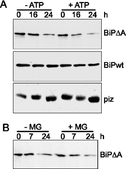
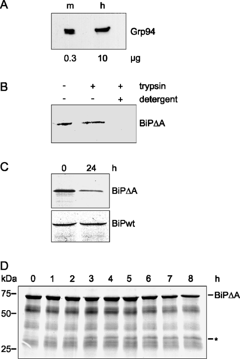
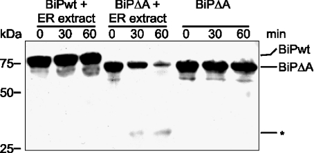
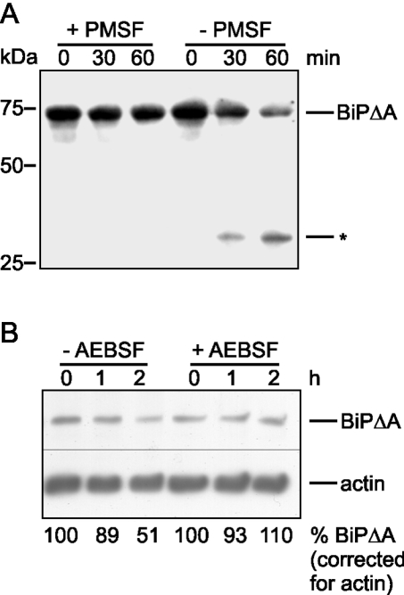
Misfolded proteins are removed from the ER (endoplasmic reticulum) by retrotranslocation to the cytosol and degradation by the ubiquitin-proteasome system in a process designated ERAD (ER-associated degradation). Analysing the turnover of a misfolded form of the ER-resident chaperone BiP (heavy-chain binding protein) (BiPDeltaA), we found that the degradation of BiPDeltaA did not follow this general ERAD pathway. In transfected cells, BiPDeltaA was degraded, although proteasome-dependent ERAD was inactivated either by proteasome inhibitors or by ATP depletion. In semi-permeabilized cells, which did not support the degradation of the proteasomal substrate alpha1-antitrypsin, the degradation of BiPDeltaA was still functional, excluding the Golgi apparatus or lysosomes as the degradative compartment. The degradation of BiPDeltaA was recapitulated in biosynthetically loaded brain microsomes and in an extract of luminal ER proteins. In contrast with proteasome-dependent ERAD, degradation fragments were detectable inside the microsomes and in the extract, and the degradation was prevented by a serine protease inhibitor. These results show that the degradation of BiPDeltaA was initiated in the ER lumen by a serine protease, and support the view that proteasome-independent ERAD pathways exist.


Figures

Figure 1
Figure 1. BiPΔA is misfolded
(A) Schematic representation of BiPwt and BiPΔA having 72 amino acids deleted in the ATPase domain. The N-terminal StrepTag epitope and the C-terminal HA epitope are indicated. pept.-bind., peptide-binding region. (B) BiPwt and BiPΔA were incubated with the indicated amounts of proteinase K (prot K) in the absence (−ATP) or presence (+ATP) of ATP. Proteins were visualized by Coomassie Blue staining.
Figure 2
Figure 2. BiPΔA is selectively degraded in CHO cells
(A) CHO cells transfected with BiPwt or BiPΔA were labelled with [35S]methionine and chased for the indicated times (in h). BiP was immunoprecipitated using an anti-HA antibody and visualized by autoradiography. u, untransfected cells. (B) Graphic representation of eight independent experiments performed as in (A). Results are means±S.D.
Figure 3
Figure 3. Degradation of BiPΔA is independent of proteasomes
(A) BiPΔA-transfected CHO cells were labelled with [35S]methionine and chased for the indicated times (in h) in the absence (−) or presence (+) of the proteasome inhibitor NLVS. BiPΔA was immunoprecipitated and visualized by autoradiography. u, untransfected cells; *, endogenous BiP sticking to the agarose beads. (B) Graphic representation of three independent experiments performed as in (A). Results are means±S.E.M. (C) BiPΔA-transfected CHO cells were labelled with [35S]methionine and chased for 8 h in the absence (−) or presence of lactacystin (LC), NLVS or MG-132 (MG). Analysis was performed as in (A).
Figure 4
Figure 4. Degradation of BiPΔA does not require ATP
BiPΔA- or piz-transfected CHO cells were labelled with [35S]methionine and chased for the indicated times (in h) in the absence (−) or presence (+) of antimycin A and 2-deoxy-D-glucose. (A) The cellular ATP content of control cells (light grey) and ATP-depleted cells (dark grey) was measured at the indicated times (in h) using a luciferase-based assay. The ATP content found in control cells at 0 h incubation was set as 100%. Results are means±S.D. (n=4). (B) BiPΔA and piz were immunoprecipitated and visualized by autoradiography. u, untransfected cells. (C) Graphical representation of three independent experiments performed as in (B). Results are means±S.E.M.
Figure 5
Figure 5. BiPΔA is degraded in semi-permeabilized cells
(A) CHO cells were transfected with BiPwt, BiPΔA or piz. After permeabilizing the plasma membrane with digitonin, the cells were chased for the indicated times (in h) in the presence of an ATP-depleting (−ATP) or ATP-regenerating (+ATP) system. The proteins were visualized by immunoblotting. (B) BiPΔA-transfected semi-permeabilized cells were chased for the indicated times (in h) in the absence (−MG) or presence (+MG) of the proteasome inhibitor MG-132. BiPΔA was visualized by immunoblotting.
Figure 6
Figure 6. BiPΔA is degraded in ER-derived microsomes
(A) Microsomes were prepared from porcine brain. The enrichment of the ER marker Grp94 in the microsomal preparation (m), as compared with brain homogenate (h), was determined by Western blot analysis. Note that different amounts of protein were loaded. (B) Microsomes were loaded with BiPΔA by in vitro translation. After re-isolation by centrifugation, the microsomes were treated with trypsin in the absence or presence of detergent. (C) BiPΔA- or BiPwt-loaded microsomes were washed with high salt to remove adsorbed cytosolic proteins, resuspended in buffer containing an ATP-depleting system and chased for the indicated times (in h). Proteins were separated by SDS/PAGE (10% gels) and visualized by autoradiography. (D) BiPΔA-loaded microsomes were chased for the indicated times (in h). Proteins were separated by SDS/PAGE (10% gels) and visualized by autoradiography. *, possible degradation intermediate of BiPΔA. Molecular masses (in kDa) are indicated.
Figure 7
Figure 7. Degradation of BiPΔA is initiated by an ER-luminal protease
Purified BiPwt or BiPΔA was incubated for the indicated times (in h) with an ER extract prepared by mild-detergent extraction of brain microsomes. BiP was visualized by Streptactin–peroxidase. *, degradation intermediate of BiPΔA. Molecular masses (in kDa) are indicated.
Figure 8
Figure 8. BiPΔA is degraded by a serine protease
(A) Recombinant BiPΔA was incubated for the indicated times (in h) with an ER extract in the presence (+) or absence (−) of PMSF and visualized by Streptactin–peroxidase. *, degradation intermediate of BiPΔA. Molecular masses (in kDa) are indicated. (B) BiPΔA-transfected CHO cells were treated with cycloheximide and chased for the indicated times (in h) in the absence (−) or presence (+) of the serine protease inhibitor AEBSF. BiPΔA and actin were visualized by immunoblotting using anti-HA and anti-actin antibodies respectively. The blots were densitometrically analysed, and the BiPΔA signal was normalized to equal actin signals.

Similar articles

Cited by

References

    1. Ellgaard L., Helenius A. Quality control in the endoplasmic reticulum. Nat. Rev. Mol. Cell Biol. 2003;4:181–191. - PubMed
    1. Trombetta E. S., Parodi A. J. Quality control and protein folding in the secretory pathway. Annu. Rev. Cell Dev. Biol. 2003;19:649–676. - PubMed
    1. Sitia R., Braakman I. Quality control in the endoplasmic reticulum protein factory. Nature (London) 2003;426:891–894. - PubMed
    1. Tsai B., Ye Y., Rapoport T. A. Retro-translocation of proteins from the endoplasmic reticulum into the cytosol. Nat. Rev. Mol. Cell Biol. 2002;3:246–255. - PubMed
    1. Kostova Z., Wolf D. H. For whom the bell tolls: protein quality control of the endoplasmic reticulum and the ubiquitin–proteasome connection. EMBO J. 2003;22:2309–2317. - PMC - PubMed

Publication types

Substances