. 2004 Dec;11(12):1729-41.
doi: 10.1016/j.chembiol.2004.11.018.

Structural basis for discriminative regulation of gene expression by adenine- and guanine-sensing mRNAs

Affiliations

Structural basis for discriminative regulation of gene expression by adenine- and guanine-sensing mRNAs

Alexander Serganov et al. Chem Biol. 2004 Dec.

Abstract

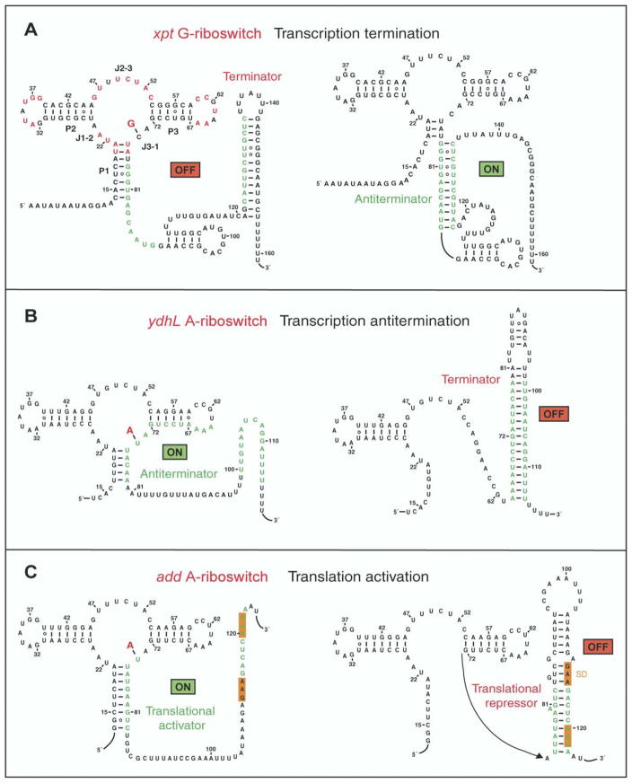
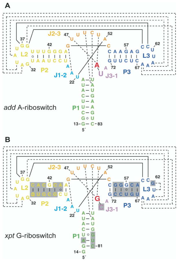
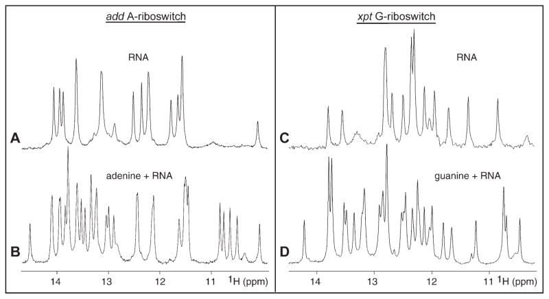
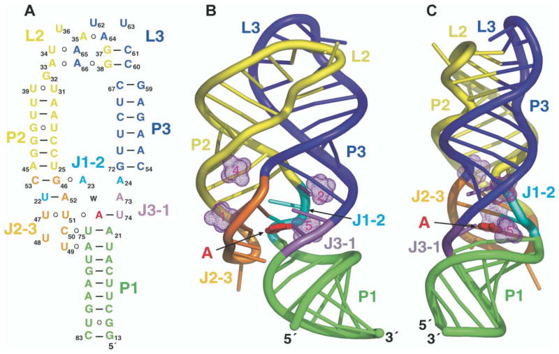
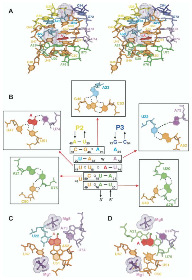
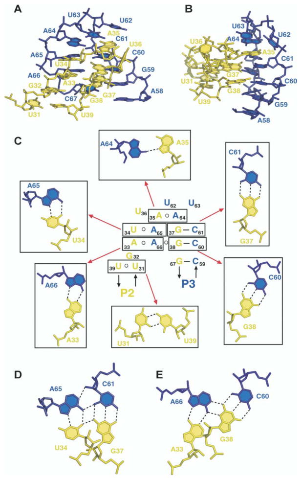
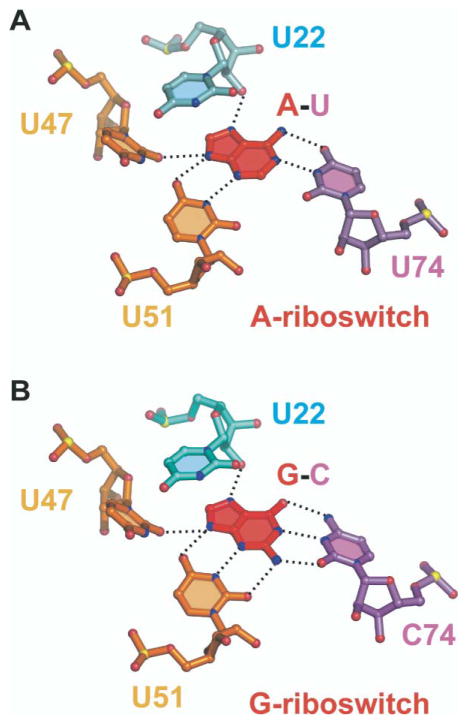
Metabolite-sensing mRNAs, or "riboswitches," specifically interact with small ligands and direct expression of the genes involved in their metabolism. Riboswitches contain sensing "aptamer" modules, capable of ligand-induced structural changes, and downstream regions, harboring expression-controlling elements. We report the crystal structures of the add A-riboswitch and xpt G-riboswitch aptamer modules that distinguish between bound adenine and guanine with exquisite specificity and modulate expression of two different sets of genes. The riboswitches form tuning fork-like architectures, in which the prongs are held in parallel through hairpin loop interactions, and the internal bubble zippers up to form the purine binding pocket. The bound purines are held by hydrogen bonding interactions involving conserved nucleotides along their entire periphery. Recognition specificity is associated with Watson-Crick pairing of the encapsulated adenine and guanine ligands with uridine and cytosine, respectively.


Figures

Figure 1
Figure 1. Mechanisms for Regulation of Gene Expression by Purine Riboswitches
(A) Schematic showing control of gene expression by the xpt G-riboswitch through transcriptional termination [23]. Highly conserved residues in the G-box of the aptamer domain are marked in red. (B) Gene expression regulation by the ydhL A-riboswitch through disruption of the terminator hairpin [24]. (C) Control of gene expression by the add A-riboswitch most likely through translational activation. The Shine-Dalgarno GAA sequence and the initiation codon are both shaded in orange.
Figure 2
Figure 2. Sequence and Stem Secondary Structures of Aptamer Domains of Purine-Sensing mRNAs
(A) V. vulnificus 71-mer add A-riboswitch; (B) B. subtilis 68-mer xpt G-riboswitch. The color-coding scheme is as follows: Stems P1, P2, and P3 are green, yellow, and blue, respectively. Loops L2 and L3 are yellow and blue, respectively. Junction-connecting segments J1-2, J2-3, and J3-1 are cyan, orange, and violet, respectively. The specificity-determining pyrimidine residue at position 74 is highlighted with a larger lettering size. The bound adenine and guanine ligands are indicated in red lettering. Tertiary pairing alignments involving Watson-Crick and noncanonical base pairs are shown by full and dashed lines, respectively. The shaded regions in (B) highlight differences between the A- and G-riboswitches.
Figure 3
Figure 3. Imino Proton NMR Spectra of Purine Binding to Aptamer Domains of Purine Riboswitches
Imino proton NMR spectra (10–15 ppm) of the 71-mer add A-riboswitch in the absence (A) and presence (B) of one equivalent of adenine and of the 69-mer (with GGC and GUC sequences on the 5′ and 3′ ends, respectively) xpt G-riboswitch in the absence (C) and presence (D) of one equivalent of guanine. The spectra were recorded in 50 mM potassium acetate (pH 6.8) at 25°C. For both riboswitches, 2 mM Mg was added to drive complex formation to completion.
Figure 4
Figure 4. Schematic and Structure of the A-Riboswitch-Adenine Complex
(A) Schematic highlighting tertiary interactions in the folded structure of the A-riboswitch-adenine complex. The color-coding is as outlined in the caption to Figure 2. (B and C) Ribbon representations (rotated by 90° along the vertical axis) of the A-riboswitch-adenine complex with the same color-coding scheme. The bound adenine is shown in red in a stick representation. Four of the five hydrated Mg cations are shown as dotted surfaces (remaining Mg is involved in packing interactions).
Figure 5
Figure 5. Details of the Tertiary Interactions within the Zippered Up Junctional Bubble in the Structure of the A-Riboswitch-Adenine Complex
(A) Stereo pair of the junction-connecting J1-2, J2-3, and J-31 segments and two junctional stem P1 base pairs that constitute the core of the complex and include the adenine binding site. The color-coding is the same as in the caption to Figure 2, with the bound adenine shown in red. (B) Schematic of the tertiary interactions involving a five-tiered arrangement of base triples. Individual base triples are boxed, and their pairing alignments (together with assignments) are shown on either side and above the schematic. Water molecule (w) is shown as a green ball. Hydrogen bonds are indicated by dotted lines. (C) Stacking of the A73•(A52-U22) triple over the shaded U51•(adenine-U74) recognition triple. (D) Stacking of the shaded U51•(adenine-U74) recognition triple over the C50•(U75-A21) triple. In (C) and (D), hydrated Mg ions, surrounded by the solvent-accessible surface in a mesh representation, are shown in ball-and-stick representation.
Figure 6
Figure 6. Details of the Tertiary Interactions between the Kissing Hairpins in the Structure of the A-Riboswitch-Adenine Complex
(A and B) Two views (rotated by 90° along the vertical axis) of the kissing interaction between loop L2, in yellow, and loop L3, in blue. (C) Schematic of the tertiary interactions between kissing hairpin loops. Individual tertiary pairs are boxed, and their pairing alignments (together with assignments) are shown around the schematic. Pairing alignment of adjacent (D) U34•A65•C61-G37 and (E) A33• A66•C60-G38 tetrads.
Figure 7
Figure 7. Recognition of Bound Purines in Purine Riboswitches
(A) Hydrogen-bonding alignments to bound adenine in the A-riboswitch. The bound adenine forms a Watson-Crick pair with U74. (B) Hydrogen-bonding alignments to bound guanine in the G-riboswitch. The bound guanine forms a Watson-Crick pair with C74. Hydrogen bonds involving 2′-OH of U22 and base edges of U47 and U51 are common to both riboswitches. Oxygen, nitrogen, and phosphorus atoms are shown, respectively, as red, blue, and yellow balls.

Comment in

References

    1. Batey RT, Rambo RP, Doudna JA. Tertiary motifs in RNA structure and folding. Angew Chem Int Ed Engl. 1999;38:2326–2343. - PubMed
    1. Hermann T, Patel DJ. Stitching together RNA tertiary architectures. J Mol Biol. 1999;294:829–849. - PubMed
    1. Doherty EA, Doudna JA. Ribozyme structure and mechanisms. Annu Rev Biophys Biomol Struct. 2001;30:457–475. - PubMed
    1. Lilley DMJ. Structure, folding and catalysis of the small nucleolytic ribozymes. Curr Opin Struct Biol. 1999;9:330–338. - PubMed
    1. Jaschke A. Artificial ribozymes and deoxyribozymes. Curr Opin Struct Biol. 2001;11:321–326. - PubMed

Publication types

MeSH terms