. 2004 Dec;168(4):1827-41.
doi: 10.1534/genetics.104.032888.

Genetic analysis of yeast Yip1p function reveals a requirement for Golgi-localized rab proteins and rab-Guanine nucleotide dissociation inhibitor

Affiliations

Genetic analysis of yeast Yip1p function reveals a requirement for Golgi-localized rab proteins and rab-Guanine nucleotide dissociation inhibitor

Catherine Z Chen et al. Genetics. 2004 Dec.

Abstract

Yip1p is the first identified Rab-interacting membrane protein and the founder member of the YIP1 family, with both orthologs and paralogs found in all eukaryotic genomes. The exact role of Yip1p is unclear; YIP1 is an essential gene and defective alleles severely disrupt membrane transport and inhibit ER vesicle budding. Yip1p has the ability to physically interact with Rab proteins and the nature of this interaction has led to suggestions that Yip1p may function in the process by which Rab proteins translocate between cytosol and membranes. In this study we have investigated the physiological requirements for Yip1p action. Yip1p function requires Rab-GDI and Rab proteins, and several mutations that abrogate Yip1p function lack Rab-interacting capability. We have previously shown that Yip1p in detergent extracts has the capability to physically interact with Rab proteins in a promiscuous manner; however, a genetic analysis that covers every yeast Rab reveals that the Rab requirement in vivo is exclusively confined to a subset of Rab proteins that are localized to the Golgi apparatus.


Figures

F<sc>igure</sc> 1.—
Figure 1.—
(A) Alignment of YIP1 orthologs identifying residues targeted for mutagenesis: sequence of Yip1p and comparison with full-length cDNAs from human YIP1 (HsYIP1), mouse YIP1 (MmYIP1), and Caenorhabditis elegans YIP1 (CeYIP1). The sequences were aligned in MegAlign (DNASTAR) using Clustal analysis with a gap-length penalty of 10. Amino acid residues are numbered according to the yeast Yip1p primary sequence. The residues on a black background exactly match the consensus sequence. (B) Mutagenized Yip1p residues are distributed in both the NH2- and the COOH-terminal domain of Yip1p. YIP1 family members share a common domain topology of a NH2-terminal hydrophilic and a COOH-terminal hydrophobic domain indicated by the Membrane Protein Explorer (MPEx) profile of Yip1p, generated with the octanol-interface scale (White and Wimley 1999; Jayasinghe et al. 2001). Yeast Yip1p residues mutagenized in this study are indicated and coded according to physiological impact designated by a symbol placed at the amino acid position of the MPEx profile of Yip1p. Table 3 contains a more detailed description of the mutant alleles. (C) Expression of a dominant-negative allele, yip1-6. Cells containing plasmids expressing yip1-6 (E76K) under the control of the copper-inducible CUP1 promoter were grown to early log phase before adjusting and adding CuSO4 to the media as indicated. Growth was assessed by OD600 turbidity measurements. At 0.5 mm Cu2w+ levels, growth ceases after 5 hr at 30° in the cells expressing yip1-6, but not in vector-only control cells.
F<sc>igure</sc> 2.—
Figure 2.—
(A) Thermosensitivity of yip1 alleles. Growth of yip1-40, yip1-42, and yip1-4 cells on YPD are compared to isogenic wild-type controls at the temperatures indicated. yip1-4 mutant cells bearing the single point mutation E70K are thermosensitive with a restrictive temperature of 34° on rich media; yip1-40 cells (E70G, K130A) and yip1-42 cells (E70A) are severely growth retarded at 37° and nonviable at 40°. (B) Interaction of Yip1p residues 70 and 130: growth phenotype of haploid cells with mutations in residue positions 70 and 130 of Yip1p. RCY1610 cells, genotype MATα ura3-52 leu2-3,112 YIP1ΔKANR [YCP50 YIP1], were transformed with pRS315 yip1 plasmids as indicated. Transformants were streaked onto 5-FOA containing media at 25° to assess growth with the mutant versions of the genes as the sole copy. All genes were expressed under the control of the endogenous promoter and terminator. The single point mutations E70K (yip1-4, pRC1992), K130A (yip1-13, pRC2398), and K130E (yip1-31, pRC2454) are viable in contrast to E70G (yip1-41, pRC2214), which is a lethal single point mutant. K130A can suppress the lethality of E70G (yip1-40, pRC2400); however, the combination of mutants in these two positions to create a charge reversal of the naturally occurring amino acids, E70K/K130E (yip1-44, pRC2452), yields a lethal phenotype, suggesting that intragenic suppression occurs with an indirect interaction of these two residues.
F<sc>igure</sc> 3.—
Figure 3.—
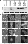
Analysis of tagged yip1 mutant alleles. (A, top) Immunoblots of total lysates of congenic log-phase GFP-tagged yip1 haploid strains, grown in minimal medium and probed with anti-GFP antibody to visualize the GFP-tagged yip1 allele. Blots were stripped with 0.2 m NaOH and reprobed with monoclonal antibody 10D7 that recognizes V-ATPase as a loading control (bottom). Negative control indicates lysates from an isogenic cell line not expressing GFP. (B) The localization of GFP-YIP1 wild-type and mutant alleles visualized in vivo by fluorescence microscopy in wild-type (RCY239) cells. (Left) GFP fluorescence; (right) DIC optics of the same field.
F<sc>igure</sc> 4.—
Figure 4.—
Synthetic lethality assay of yip1 alleles with Rab-encoding genes and genes regulating Rab protein function. (A) Synthetic lethality plate assay: strains carrying thermosensitive mutations of Rab protein ORFs and various genes involved in Rab protein function and a deletion of YIP1 together with wild-type YIP1 on a URA3 plasmid were transformed with vector-only control (pRS315), wild-type YIP1 control (pRC1838B), and the thermosensitive allele yip1-4 (pRC1992). All transformants were assessed by plasmid shuffling on 5-FOA at 25°. Plates bearing vps21Δ sec4-8, ypt11Δ double mutants with yip1-4 are shown as examples where no genetic interaction was observed. Similarly, ypt31Δ and ypt32A134D with yip1-4 double mutants are shown as examples of complete lethality between two mutants. (B) Testing multiple alleles for synthetic lethality: isogenic strains bearing various yip1 conditional lethal alleles as indicated were tested for genetic interactions at 25° with the temperature-sensitive gene ypt1A136D. Wild-type YIP1 and empty vector are included as a control for positive and negative growth, respectively.
F<sc>igure</sc> 5.—
Figure 5.—
Y2H interactions between yip1 mutant alleles and Rab-interacting partners. Pairs of constructs were coexpressed in the reporter strain Y190 and β-galactosidase activity (arbitrary units) in the resulting transformants was measured as described in materials and methods. The protein “bait” constructs were tested against “prey” constructs of Yip1p mutant alleles, Yip1p, or vector-only (pAS2-1) controls as indicated on the y-axis. Shaded bars, yip1 alleles containing mutations in the NH2 terminus; solid bars, COOH-terminal alleles. Plasmid constructs are listed for Ypt1p interactions; the same constructs were used for all other experiments.
F<sc>igure</sc> 6.—
Figure 6.—
yip1 membranes display a defect in COPII vesicle budding in vitro. Washed semi-intact cells containing [35S]gpαf were prepared from wild-type and yip1-40 (A) or yip1-42 (B) strains. Semi-intact cells were incubated with buffer (NA) or a reconstitution mixture (Recon), containing COPII, Uso1p, LMA1, and an ATP regeneration system, to measure ER-to-Golgi transport. After 75 min at 23°, the amount of Golgi-modified [35S]gpαf was measured to determine transport efficiency. (C) Semi-intact cells from wild-type and yip1-42 mutant strains were prepared as in B and incubated with COPII proteins to measure vesicle budding efficiency. After 30 min at 23°, freely diffusible vesicles containing [35S]gpαf were separated from semi-intact cell membranes by centrifugation at 18,000 × g and [35S]gpαf quantified by ConA precipitation.
F<sc>igure</sc> 7.—
Figure 7.—
Two models of Yip1p function. Genetic analysis shows that Yip1p “buffers” (Hartman et al. 2001) the action of COPII genes in ER vesicle biogenesis and genes encoding Golgi Rab proteins. Although the mechanisms that underlie these genetic relationships are not known, these data suggest two models: (A) Yip1p operates independently in ER vesicle biogenesis and with Golgi Rab proteins, and (B) Yip1p function links ER vesicle biogenesis with the action of Golgi Rab proteins.

References

    1. Aguilera, A., 1994. Formamide sensitivity: a novel conditional phenotype in yeast. Genetics 136: 87–91. - PMC - PubMed
    1. Allan, B. B., B. D. Moyer and W. E. Balch, 2000. Rab1 recruitment of p115 into a cis-SNARE complex: programming budding COPII vesicles for fusion. Science 289: 444–448. - PubMed
    1. Alory, C., and W. E. Balch, 2001. Organization of the Rab-GDI/CHM superfamily: the functional basis for choroideremia disease. Traffic 2: 532–543. - PubMed
    1. Baker, D., L. Hicke, M. Rexach, M. Schleyer and R. Schekman, 1988. Reconstitution of SEC gene product-dependent intercompartmental protein transport. Cell 54: 335–344. - PubMed
    1. Barlowe, C., 1997. Coupled ER to Golgi transport reconstituted with purified cytosolic proteins. J. Cell Biol. 139: 1097–1108. - PMC - PubMed

Publication types

MeSH terms

Substances