. 2004 Dec 20;200(12):1613-22.
doi: 10.1084/jem.20041395.

Age-related defects in CD4 T cell cognate helper function lead to reductions in humoral responses

Affiliations

Age-related defects in CD4 T cell cognate helper function lead to reductions in humoral responses

Sheri M Eaton et al. J Exp Med. .

Abstract

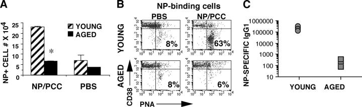
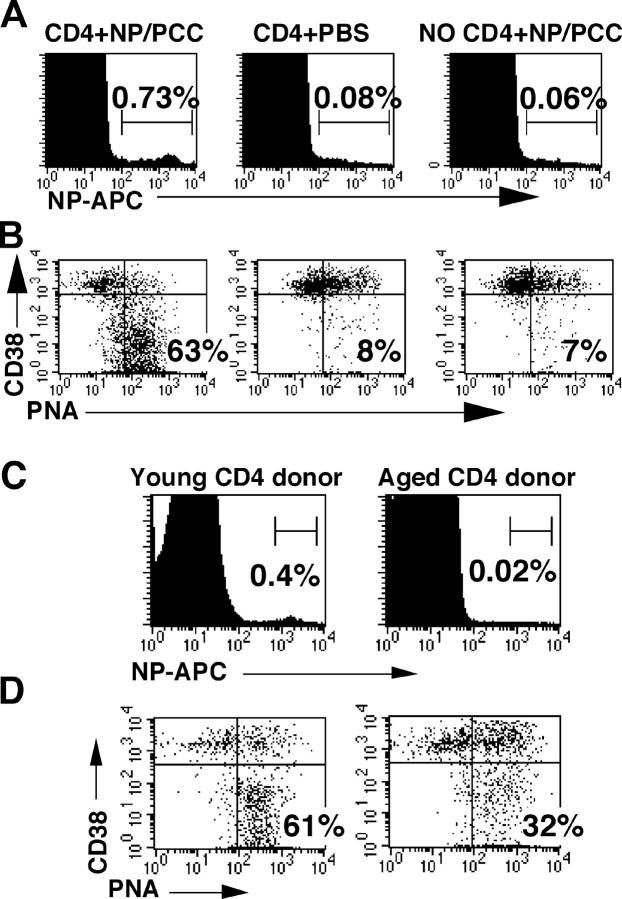
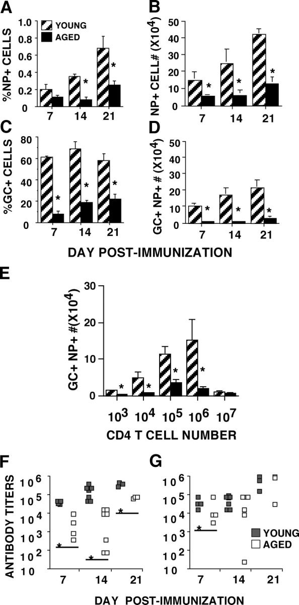
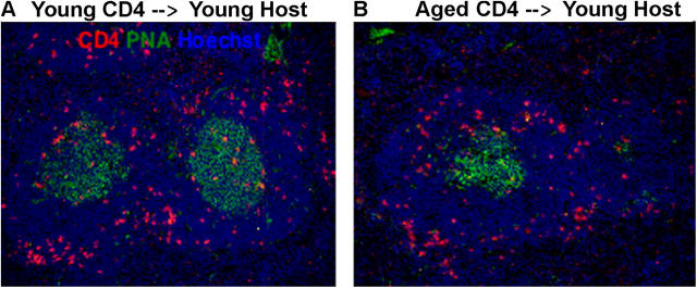
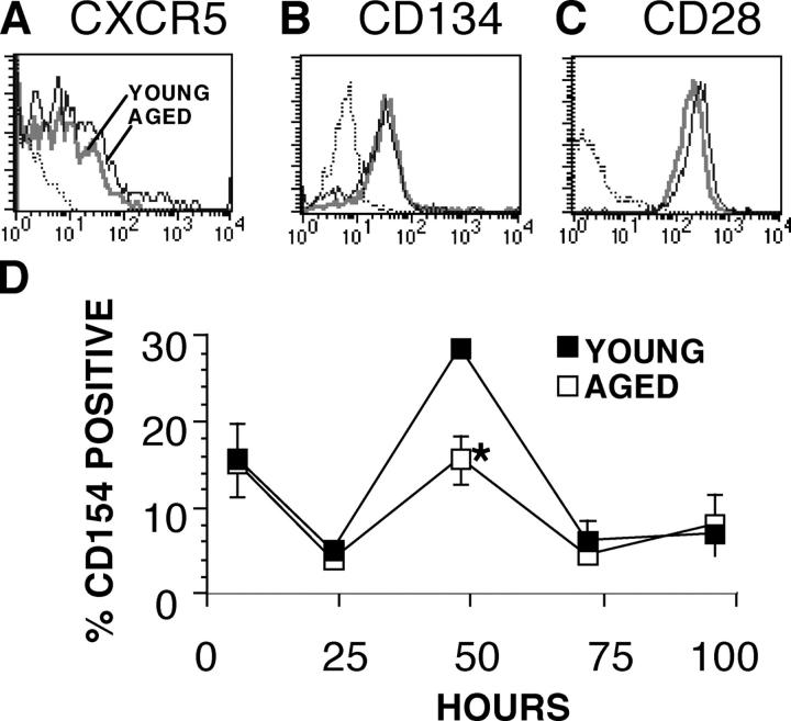
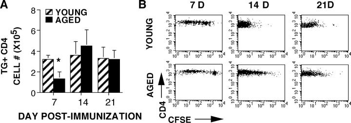
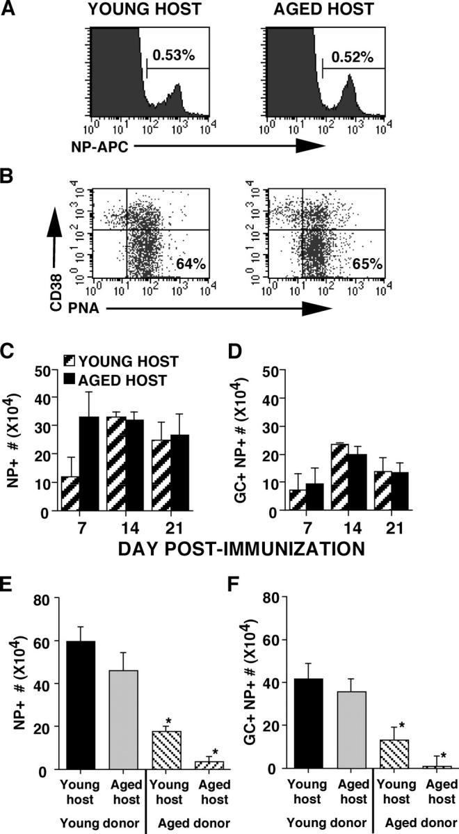
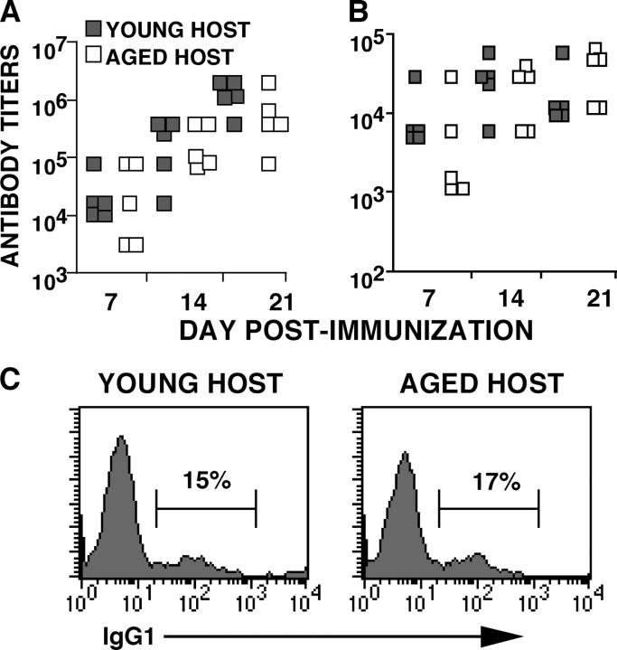
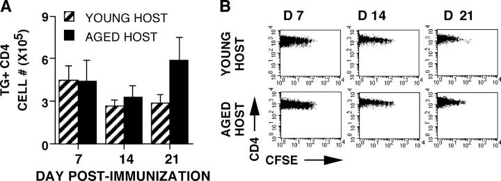
With increasing age, the ability to produce protective antibodies in response to immunization declines, leading to a reduced efficacy of vaccination in the elderly. To examine the effect of age on the cognate function of CD4 T cells, we have used a novel adoptive transfer model that allows us to compare identical numbers of antigen-specific naive T cells from young and aged TCR transgenic (Tg) donors. Upon transfer of aged donor CD4 T cells to young hosts, there was significantly reduced expansion and germinal center (GC) differentiation of the antigen-specific B cell population after immunization. This reduced cognate helper function was seen at all time points and over a wide range of donor cell numbers. In hosts receiving aged CD4 cells, there were also dramatically lower levels of antigen-specific IgG. These age-related defects were not due to defects in migration of the aged CD4 T cells, but may be attributable to reduced CD154 (CD40L) expression. Furthermore, we found that there was no difference in B cell expansion and differentiation or in IgG production when young CD4 T cells were transferred to young or aged hosts. Our results show that, in this model, age-related reductions in the cognate helper function of CD4 T cells contribute significantly to defects in humoral responses observed in aged individuals.


Figures

Figure 1.
Figure 1.
Decreased B cell expansion and GC formation in aged mice. Young (2 mo) and aged (23 mo) nontransgenic B10.Br mice were immunized i.p. with NP-PCC/alum (200 μg) or PBS/alum. On day 14, spleens were harvested and stained with NP-APC to detect NP-binding cells by flow cytometry. (A) Bar graph shows the total number of NP-binding cells in young (striped bars) and aged (solid bars) groups (mean ± SE). (B) Representative flow cytometry dot plots gated on NP-binding cells showing CD38 and PNA expression. (C) NP-specific serum IgG1 concentrations in each of the young and aged immunized mice. *P < 0.05.
Figure 2.
Figure 2.
Adoptive transfer model to study in vivo cognate helper function. (A) Young AND Tg CD4 cells (106) were adoptively transferred i.v. to young CD4KO hosts. Control hosts received no CD4 cells. Hosts were then immunized i.p. with 200 μg NP/PCC or PBS in alum. On day 14, NP-specific expansion was examined by staining splenocytes with NP-APC. The percent of NP-binding cells for each condition is shown in the flow cytometry histograms. (B) Dot plots showing PNA versus CD38 staining are gated on NP-binding cells in A. Percentages indicate GC phenotype B cells (CD38loPNAhi). (C) Flow cytometry histograms showing expansion of NP-specific cells on day 14 after immunization with NP-PCC and transfer of young or aged donor CD4 T cells. (D) Dot plots are gated on NP-binding cells in C; percentages indicate the GC+ phenotype population.
Figure 3.
Figure 3.
Reduced cognate helper function of Tg CD4 T cells from aged mice. Young (striped bars) and aged (solid bars) AND Tg CD4 cells (106) were adoptively transferred i.v. to young CD4KO hosts. Hosts were then immunized i.p. with 200 μg NP/PCC in alum. On days 7, 14, and 21 the (A) percentages and (B) numbers of NP-binding cells were determined by flow cytometry following staining with NP-APC (mean ± SE). On days 7, 14, and 21 the (C) percentages and (D) numbers of GC phenotype (CD38loPNAhi) cells within the NP-binding population was determined by flow cytometry (mean ± SE). (E) Numbers of GC phenotype (CD38loPNAhi) NP-binding cells per spleen on day 14 upon titration of donor T cell numbers. The numbers of young and aged donor cells were titrated from 103 to 107 per host. Data shown is mean ± SE. (F) NP-specific serum IgG1 titers in NP-PCC immunized hosts receiving young (shaded squares) or aged (open squares) Tg CD4 T cells on days 7, 14, and 21 after immunization. (G) NP-specific serum IgM titers in NP-PCC immunized hosts receiving young (shaded squares) or aged (open squares) Tg CD4 T cells on days 7, 14, and 21 after immunization. For all experiments: *P < 0.05.
Figure 4.
Figure 4.
Young and aged Tg CD4 T cells migrate similarly in young hosts after immunization. On day 14 after immunization with NP-PCC/alum and transfer of young and aged donor T cells into young CD4KO hosts, the migration of donor cells into follicles was examined by fluorescence microscopy. Frozen sections were cut from spleens of young hosts receiving (A) young or (B) aged donor Tg CD4 T cells and stained with anti-CD4 (red), PNA (green), and Hoechst (blue).
Figure 5.
Figure 5.
Phenotype of young and aged CD4 T cells. (A) Young (bold gray line) and aged (thin black line) donor T cells from the experiment in Fig. 4 (day 14 after immunization) were prepared for flow cytometry and stained for expression of CD4, Vβ3, and CXCR5. The flow cytometry dot plots are gated on CD4+Vβ3+ T cells and show the percent positive for CXCR5 expression. (B) Naive young and aged Tg CD4 T cells were stimulated with Ag/APC for 4 d. Effector populations were harvested and stained for CD4, Vβ3, and CD134 expression; histogram is gated on Vβ3+CD4+ cells. (C) 4 d young and aged Tg effectors were also stained for CD4, Vβ3, and CD28 expression; histogram is gated on Vβ3+CD4+ cells. (D) Young (filled squares) and aged (open squares) Tg CD4 T cells were stimulated in vitro with Ag/APC. At 6, 24, 48, 72, and 96 h, cultures were harvested, stained for CD4, Vβ3, and CD154 expression and analyzed by flow cytometry. T cell populations were gated on CD4+ Vβ3+ T cells and the percent CD154 positive in young and aged populations were determined. Graph shows the mean ± SE; *P < 0.05. Representative of three experiments.
Figure 6.
Figure 6.
Recovery of young and aged donor T cells in young hosts. Young and aged Tg CD4 T cells were CFSE labeled and transferred to young CD4KO hosts that were then immunized with NP-PCC. (A) The number of young (striped bars) and aged (solid bars) donor T cells was determined by flow cytometry analysis on days 7, 14, and 21. Data shows mean ± SE. (B) Flow cytometry dot plots, gated on Vβ3+CD4+ donor cells, showing CFSE profiles of young and aged cells on days 7, 14, and 21 after immunization.
Figure 7.
Figure 7.
Cognate helper function of young donor T cells is similar in young and aged hosts. Young Tg CD4 T cells (106) were adoptively transferred i.v. to young or aged CD4KO hosts. Hosts were then immunized i.p. with 200 μg NP/PCC or PBS in alum. (A) On day 14, the percentage of NP+ cells in young and aged hosts was determined by staining splenocytes with NP-APC. Representative flow cytometry histograms are shown. (B) Flow cytometry dot plots are gated on NP+ cells from A. Percentages indicate GC phenotype cells (CD38lowPNAhi) in young and aged hosts. (C) On days 7, 14, and 21 the numbers of NP-binding cells in young (striped bars) and aged (solid bars) hosts were determined by flow cytometry after staining with NP-APC. (D) On days 7, 14, and 21 the numbers of GC phenotype (CD38loPNAhi) cells within the NP-binding population in young (striped) and aged (solid) hosts was determined by flow cytometry. (E) Young or aged donor Tg CD4 T cells (106) were transferred into young or aged CD4KO hosts, which were immunized with NP-PCC. On day 14, the number of NP-binding cells was determined by flow cytometry. (F) The number of GC phenotype NP-binding cells was also determined on day 14 by flow cytometry. For all, data shown is mean ± SE; *P < 0.05 when aged donors compared with young donors.
Figure 8.
Figure 8.
Antibody production in young and aged hosts. Young and aged CD4KO hosts were given young donor CD4 T cells and immunized as described in the previous figure. Serum was collected on days 7, 14, and 21 after immunization. (A) NP-specific IgG1 titers were determined by ELISA from serum of young (filled squares) and aged (open squares) hosts. (B) NP-specific IgM titers were determined by ELISA from serum of young (filled squares) and aged (open squares) hosts. (C) Splenocytes from young and aged hosts were stained for NP binding and surface IgG1 expression on day 14 after immunization. Representative flow cytometry histograms are gated on NP+ splenocytes; percentages indicate the positive staining for IgG1.
Figure 9.
Figure 9.
Recovery of young donor T cells in young and aged hosts. Young Tg CD4 T cells were CFSE labeled and transferred to young and aged CD4KO hosts that were then immunized with NP-PCC. (A) The number of young donor cells in young (striped bars) and aged (solid bars) hosts was determined by flow cytometry analysis on days 7, 14, and 21. (B) Flow cytometry dot plots, gated on Vβ3+CD4+ donor cells, showing CFSE profiles from young and aged hosts on days 7, 14, and 21 after immunization.

References

    1. Kosco, M.H., G.F. Burton, Z.F. Kapasi, A.K. Szakal, and J.G. Tew. 1989. Antibody-forming cell induction during an early phase of germinal centre development and its delay with ageing. Immunology. 68:312–318. - PMC - PubMed
    1. Goidl, E.A., J.B. Innes, and M.E. Weksler. 1976. Immunological studies of aging. II. Loss of IgG and high avidity plaque- forming cells and increased suppressor cell activity in aging mice. J. Exp. Med. 144:1037–1048. - PMC - PubMed
    1. Cook, J.M., N. Gualde, L. Hessel, M. Mounier, J.P. Michel, F. Denis, and M.H. Ratinaud. 1987. Alterations in the human immune response to the hepatitis B vaccine among the elderly. Cell. Immunol. 109:89–96. - PubMed
    1. Musher, D.M., A.J. Chapman, A. Goree, S. Jonsson, D. Briles, and R.E. Baughn. 1986. Natural and vaccine-related immunity to Streptococcus pneumoniae. J. Infect. Dis. 154:245–256. - PubMed
    1. Phair, J., A. Kauffman, A. Bjornson, L. Adams, and C. Linnemann. 1978. Failure to respond to influenza vaccine in the aged: correlation with B-cell number and function. J. Lab. Clin. Med. 92:822–828. - PubMed

Publication types

MeSH terms