. 2004 Dec 20;200(12):1647-55.
doi: 10.1084/jem.20041215.

Anthrolysin O and other gram-positive cytolysins are toll-like receptor 4 agonists

Affiliations

Anthrolysin O and other gram-positive cytolysins are toll-like receptor 4 agonists

Jin Mo Park et al. J Exp Med. .

Abstract

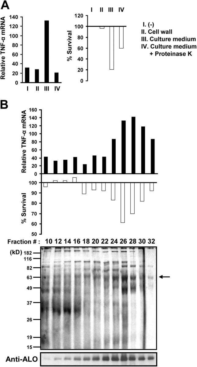
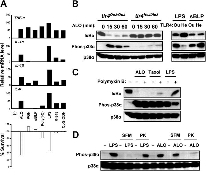
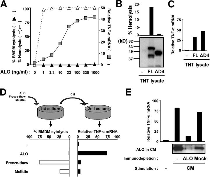
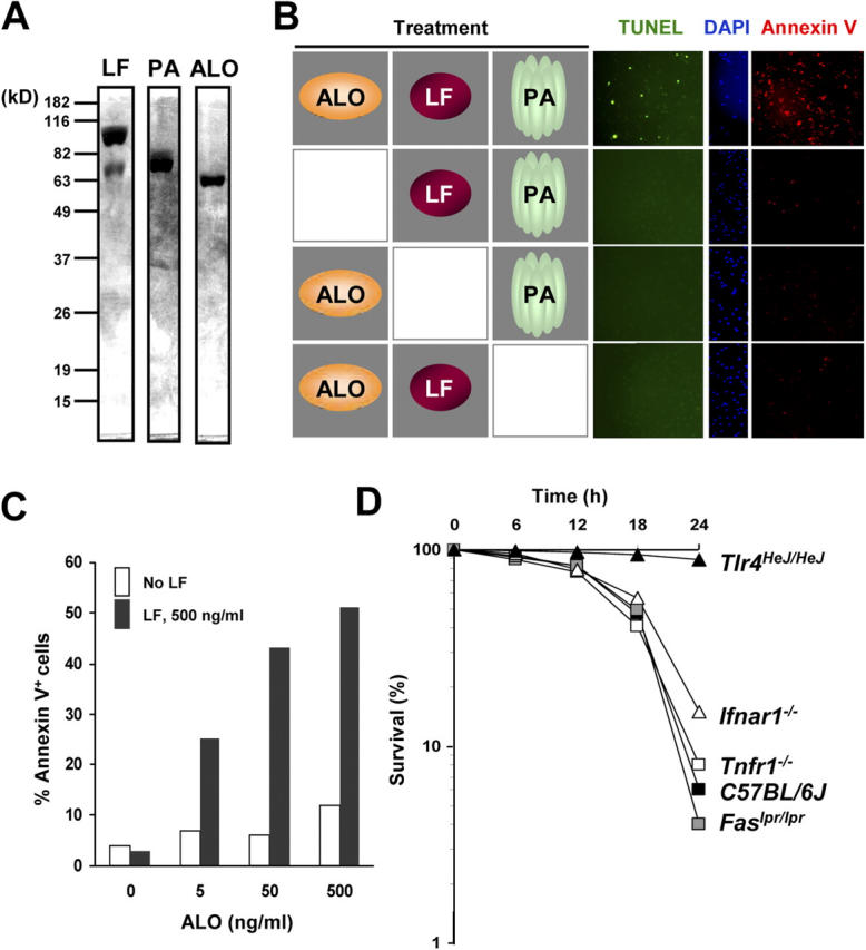
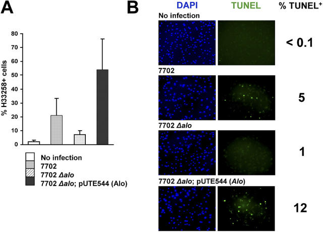
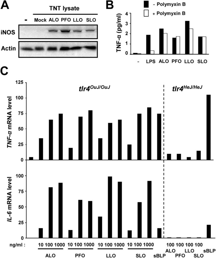
Exposure of bone marrow-derived macrophages (BMDMs) to low concentrations of Bacillus anthracis lethal toxin (LT), whose catalytic subunit is lethal factor (LF), results in induction of a robust apoptotic response dependent on activation of Toll-like receptor (TLR)4. A similar TLR4-dependent apoptotic response is observed when BMDMs are infected with live B. anthracis (Sterne strain). However, TLR4 is considered to be a specific signaling receptor for lipopolysaccharide (LPS), a typical product of gram-negative bacteria, whereas B. anthracis is gram-positive. To understand how B. anthracis can activate TLR4, we analyzed its culture supernatants and found them to contain a potent TLR4-stimulating activity that can also induce apoptosis in macrophages in which the antiapoptotic p38 MAP kinase (whose activation is prevented by LF) was inhibited. Purification of this activity suggested it consists of anthrolysin O (ALO), a member of the cholesterol-dependent cytolysin (CDC) family. We show that recombinant ALO can activate TLR4 in a manner independent of LPS contamination and, together with LT, can induce macrophage apoptosis. We also provide genetic evidence that ALO is required for induction of macrophage apoptosis in response to infection with live B. anthracis and that other CDC family members share the ability to activate TLR4.


Figures

Figure 1.
Figure 1.
Identification of a macrophage-stimulating activity in B. anthracis culture supernatant. (A) C57BL/6J BMDMs were left untreated (I), treated with a B. anthracis cell wall preparation (II), or with B. anthracis culture supernatant before (III) and after (IV) proteinase K pretreatment. After 4 h, total RNA was isolated, and relative expression of TNF-α mRNA (black bars) was determined by real-time PCR. In a separate experiment, BMDMs were preincubated with 10 μM SB202190 for 2 h, and treated as described before. After 24 h, cell viability (white bars) was measured by the MTT assay. (B) Partially purified B. anthracis culture supernatant was fractionated on a phenyl-sepharose column. Column fractions were tested for their ability to induce TNF-α expression (top) or macrophage apoptosis (middle) as described before. The protein composition and ALO-content of the column fractions were examined by SDS-PAGE and silver staining and immunoblotting with an anti-ALO antibody (bottom).
Figure 2.
Figure 2.
Macrophage activation by ALO is TLR4 dependent. (A) BMDMs were left untreated (−), or treated with 50 ng/ml recombinant ALO, or other TLR agonists, including 10 μg/ml peptidoglycan (PGN), 1 μg/ml synthetic bacterial lipopeptide (sBLP; Pam3CSK), 10 μg/ml poly(I-C), 100 ng/ml LPS, 1 μM R-848, or 1 μM CpG oligodeoxynucleotide (CpG ODN). Total RNA was analyzed as in Fig. 1 A for expression of different cytokine mRNAs (black bars). BMDMs were also preincubated with 10 μM SB202190 for 2 h and treated as before. Cell viability was measured by the MTT assay (white bars). (B) BMDMs from C3H/OuJ (tlr4 OuJ/OuJ; Ou) or C3H/HeJ (tlr4 HeJ/HeJ; He) mice were incubated with 50 ng/ml ALO, 100 ng/ml LPS, or 1 μg/ml sBLP. At the indicated time points, cell lysates were prepared and analyzed by immunoblotting with the antibodies indicated on the left for IκBα degradation and p38 activation (appearance of phosphorylated p38α). (C) BMDMs (C57BL/6J) were preincubated with or without 25 μg/ml polymyxin B for 1 h and treated with 50 ng/ml ALO, 10 μM taxol, or 100 ng/ml LPS. After 15 min, cell lysates were prepared and analyzed as in B. An IκBα signal indicates no IKK activation, whereas a phospho-p38α signal indicates p38 activation. (D) BMDMs (C57BL/6J) were incubated with LPS or ALO in either serum-containing or serum-free media (SFM) before or after pretreatment with proteinase K (PK). After 15 min, cell lysates were prepared and analyzed as in B for p38 activation.
Figure 3.
Figure 3.
ALO activates TLR4 independently of its cytolytic activity. (A) The levels of hemolysis, macrophage cytolysis (permeability to Hoechst 33258), and TNF-α mRNA induction were determined after treatment of either erythrocytes or macrophages with the indicated ALO concentrations. (B) Reticulocyte lysates programmed with either an empty backbone vector (−, pRSET-A), T7 expression vectors for full-length (FL) ALO, or the ΔD4 derivative, lacking amino acids 409–512, were analyzed by immunoblotting with an ALO-specific antibody. The hemolytic activity of the reticulocyte lysates was measured and is shown above the immunoblot. (C) BMDMs (C57BL/6J) were incubated with reticulocyte lysates prepared as in B. After 4 h, total RNA was isolated, and relative expression of TNF-α mRNA (black bars) was determined by real-time PCR. (D) BMDMs (C57BL/6J) were treated with 100 ng/ml ALO or 100 μg/ml melittin, or subjected to two cycles of freezing at −80°C and thawing at 37°C. After 4 h, the level of cell permeabilization was analyzed by staining with H33258 (left). The conditioned media (CM) collected after the first treatment were applied to fresh BMDMs and TNF-α mRNA induction was analyzed after 4 h by real-time PCR (right). (E) CM collected from ALO-treated cells was depleted with anti-ALO or preimmune serum (Mock). The level of ALO in the immunodepleted CM was analyzed by immunoblotting and the TNF-α–inducing activity of the immunodepleted CM was analyzed as in D.
Figure 4.
Figure 4.
Reconstitution of macrophage apoptosis with recombinant LF, PA, and ALO proteins. (A) Recombinant LF, PA, and ALO proteins were analyzed by SDS-PAGE and Coomassie blue staining. (B) BMDMs (C57BL/6J) were treated with different combinations of 500 ng/ml recombinant LF, 1 μg/ml PA, and 50 ng/ml ALO as indicated. After 24 h, the cells were analyzed by TUNEL, DAPI, and annexin V staining. (C) BMDMs were treated with 1 μg/ml PA and the indicated amounts of ALO in the absence (white bars) or presence (black bars) of 500 ng/ml LF. The number of apoptotic cells was determined by annexin V staining. (D) BMDMs from wild type (C57BL/6J), TLR4 mutant (Tlr4 HeJ/HeJ), IFNR1 knockout (Ifnar1 −/−), TNFR1 knockout (Tnfr1 −/−), and Fas mutant (Fas lpr/lpr) mice were preincubated with 10 μM SB202190 and treated with 50 ng/ml ALO. At different time points, cell viability was determined by Hoechst 33258 staining. Cells that were not stained were counted as viable cells.
Figure 5.
Figure 5.
Requirement for ALO in B. anthracis–induced macrophage apoptosis. (A) BMDMs (C57BL/6J) were infected with wild-type B. anthracis (7702), alo mutant (7702Δalo), or alo mutant complemented with the alo gene (7702Δalo; pUTE544[Alo]) at a multiplicity of infection of 0.1. After 6 h, cell death was measured by H33258 staining. (B) BMDMs were infected with B. anthracis as in A. After 12 h, cells were stained with DAPI (blue) or TUNEL (green).
Figure 6.
Figure 6.
TLR4 activation by ALO and other CDCs of gram-positive bacteria. (A) BMDMs (C57BL/6J) were incubated with reticulocyte lysates programmed with either an empty backbone vector (Mock; pRSET-A), or T7 expression vectors for ALO, perfringolysin O (PFO), listeriolysin O (LLO), or streptolysin O (SLO). After 12 h, cell lysates were prepared and analyzed by immunoblotting for iNOS induction. (B) BMDMs (C57BL/6J) were preincubated with or without 25 μg/ml polymyxin B for 1 h and treated with100 ng/ml LPS or 50 ng/ml CDC proteins. After 12 h, culture supernatants were collected and examined for TNF-α secretion by ELISA. (C) BMDMs from C3H/OuJ (tlr4 OuJ/OuJ) or C3H/HeJ (tlr4 He HeJ/HeJ) mice were stimulated with the indicated amounts of recombinant ALO, PFO, LLO, and SLO, or sBLP (1 μg/ml). After 4 h, total RNA was isolated, and the levels of TNF-α and IL-6 mRNA were measured by real-time PCR.

References

    1. Dixon, T.C., M. Meselson, J. Guillemin, and P.C. Hanna. 1999. Anthrax. N. Engl. J. Med. 341:815–826. - PubMed
    1. Collier, R.J., and J.A. Young. 2003. Anthrax toxin. Annu. Rev. Cell Dev. Biol. 19:45–70. - PubMed
    1. Moayeri, M., and S.H. Leppla. 2004. The roles of anthrax toxin in pathogenesis. Curr. Opin. Microbiol. 7:19–24. - PubMed
    1. Bradley, K.A., J. Mogridge, M. Mourez, R.J. Collier, and J.A. Young. 2001. Identification of the cellular receptor for anthrax toxin. Nature. 414:225–229. - PubMed
    1. Scobie, H.M., G.J. Rainey, K.A. Bradley, and J.A. Young. 2003. Human capillary morphogenesis protein 2 functions as an anthrax toxin receptor. Proc. Natl. Acad. Sci. USA. 100:5170–5174. - PMC - PubMed

Publication types

MeSH terms