. 2004 Dec 20;200(12):1673-9.
doi: 10.1084/jem.20041545.

Cadherin-11 provides specific cellular adhesion between fibroblast-like synoviocytes

Affiliations

Cadherin-11 provides specific cellular adhesion between fibroblast-like synoviocytes

Xavier Valencia et al. J Exp Med. .

Abstract

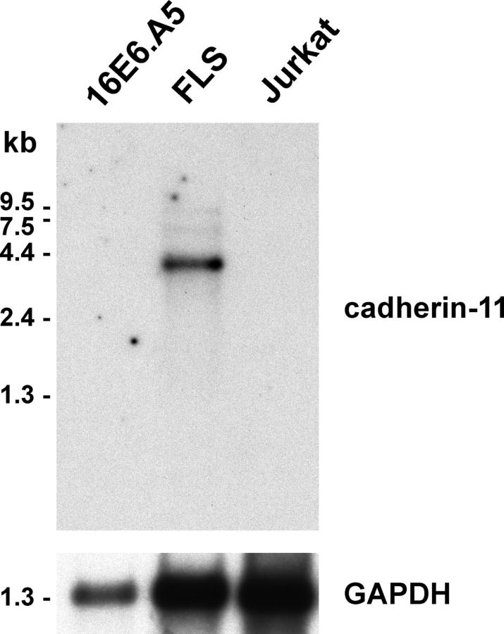
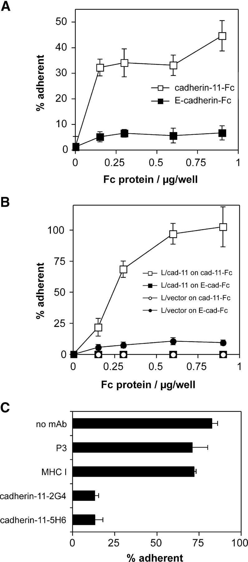
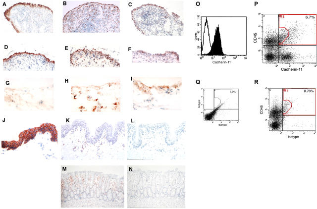
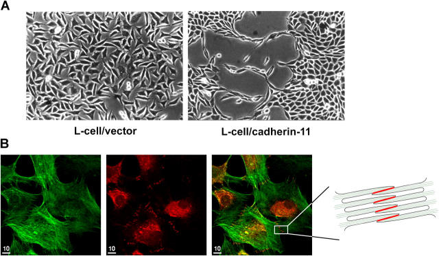
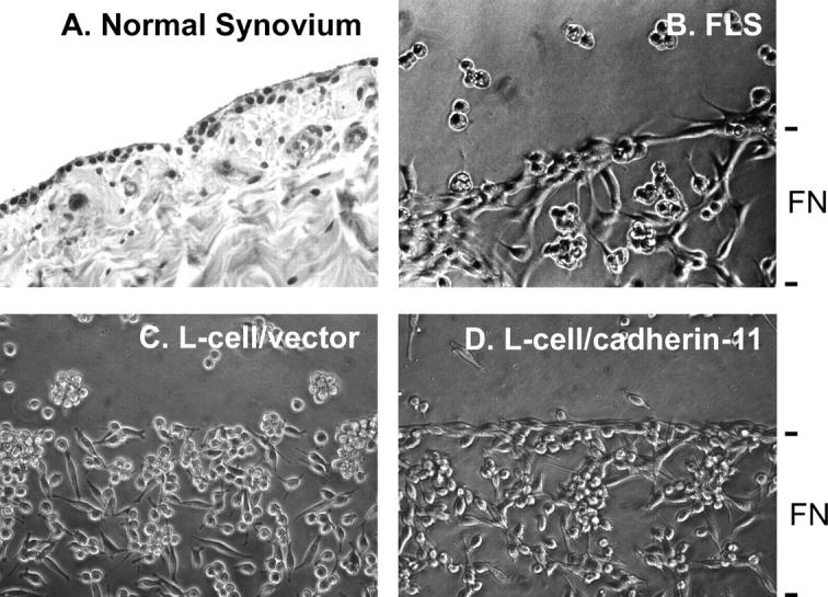
Cadherins are integral membrane proteins expressed in tissue-restricted patterns that mediate homophilic intercellular adhesion. During development, they orchestrate tissue morphogenesis and, in the adult, they determine tissue integrity and architecture. The synovial lining is a condensation of fibroblast-like synoviocytes (FLS) and macrophages one to three cells thick. These cells are embedded within the extracellular matrix, but the structure is neither an epithelium nor an endothelium. Previously, the basis for organization of the synovium into a tissue was unknown. Here, we cloned cadherin-11 from human rheumatoid arthritis (RA)-derived FLS. We developed L cell transfectants expressing cadherin-11, cadherin-11 fusion proteins, and anti-cadherin-11 mAb. Cadherin-11 was found to be expressed mainly in the synovial lining by immunohistologic staining of human synovium. FLS adhered to cadherin-11-Fc, and transfection of cadherin-11 conferred the formation of tissue-like sheets and lining-like structures upon fibroblasts in vitro. These findings support a key role for cadherin-11 in the specific adhesion of FLS and in synovial tissue organization and behavior in health and RA.


Figures

Figure 1.
Figure 1.
Northern analysis of cadherin-11 expression. (A) Northern blot of mRNA (5 μg per lane) from the 16E6.A5 breast epithelial cell line, human RA-derived cultured FLS, and Jurkat T cells was hybridized with a 32P-labeled cadherin-11 (top) or control GAPDH probe (bottom).
Figure 2.
Figure 2.
Cadherin-11–mediated adhesion of FLS and L cell transfectants. (A) Adhesion of cultured FLS derived from RA synovium to cadherin-11–Fc. Purified cadherin-11–Fc (open squares) or E-cadherin–Fc (closed squares) were immobilized on microtiter plate wells coated with polyclonal goat anti–human IgG antibody and blocked with BSA. The adhesion of RA-derived cultured FLS was determined in triplicate as described in Materials and Methods. (B) Specific adhesion of L cells to cadherin-11–Fc is conferred by cadherin-11 transfection. The assay was conducted as in A using control L cells (L/vector) or cadherin-11–transfected L cells (L/cad-11) and microtiter plates directly coated with the indicated concentrations of Fc proteins (cadherin-11–Fc or control E-cadherin–Fc). (C) Inhibition of adhesion of the L cell/cadherin-11 line to cadherin-11–Fc by anti–cadherin-11 antibodies. The assay was performed as in B but antibodies were preincubated with the cells for 10 min on ice. All the mAb were purified and used at 10 μg/ml and all mAb were mouse IgG1, except anti–mouse MHC-I (mouse IgG2a). The results are expressed as the mean percentage of cells that were adherent ±1 SD (n = 3).
Figure 3.
Figure 3.
Cadherin-11 expression on FLS in the synovium. (A–N) Immunohistochemical analyses of frozen human tissue from RA synovium (A–C), osteoarthritis (OA) synovium (D–F), normal synovium (G–I), skin (J–L), and colon (M and N) stained with anti-CD55 (A and D), anti-CD68 (B, E, H, K, and M), anti–cadherin-11 (C, F, I, L, and N), IgG1 control (G), or anti–E-cadherin (J) are shown. Note the prominent cadherin-11 reactivity in the synovial lining that was similar to CD55 reactivity and the absence of detectable cadherin-11 reactivity in skin and colonic sections. Note also the distinct difference in reactivity pattern for the macrophage marker CD68. Magnification, 200. (O–R) Flow cytometry of FLS. Flow cytometry of passage three RA-derived FLS revealed that, after in vitro culture, essentially all express cadherin-11 (O), whereas nonfibroblast lineages do not propagate. Flow cytometric analyses of freshly disaggregated RA synovial cells, gated to exclude small particulate material by forward scatter, show separate subpopulations stained by anti–cadherin-11–biotin/streptavidin-Cychrome and the bone marrow lineage marker CD45-FITC (P), compared with IgG1 controls (Q) or to synovial cells stained with CD45-FITC and isotype control (R).
Figure 4.
Figure 4.
Cadherin-11 mediates the formation of intercellular junctions, tissue sheet-like and lining-like cellular organization. (A) Cadherin-11 mediates tissue–sheet formation. Equal numbers of L cells were plated on culture dishes in conventional media. After 4 d, L cells transfected with vector alone were randomly organized without specific cell–cell interactions. In contrast, cadherin-11–transfected L cells formed extensive and intimate contacts along their surfaces and condensed together to form a continuous sheet of cells resembling a tissue-like structure. (B) Formation of cadherin-11 mediated intercellular junctions. FLS were plated at intermediate density and, after overnight culture, subjected to immunofluorescence using phalloidin to label F-actin (green, left) and anti–cadherin-11 3H10 (red, middle). A merged image is shown in at right. F-actin staining demonstrated numerous filopodia that interdigitated with those from neighboring cells. At the cell periphery, the anti–cadherin-11-3H10 mAb–labeled intercellular junctions resulted in a row of short parallel lines (middle, right, and schematic representation). Bars, 10 μm.
Figure 5.
Figure 5.
Cadherin-11 mediates lining-like formation by rheumatoid FLS and L cell transfectants. (A) Normal synovium stained with hematoxylin and eosin. The synovial lining is a thin one to three cell layer of cells at the surface of the sublining composed of the loose connective tissue, containing scattered fibroblasts, blood vessels, and ECM. (B) In vitro synovial lining formation. Discrete regions of tissue culture flasks were coated with fibronectin, followed by blocking of the entire surface with BSA. RA-derived human FLS were plated and cultured under serum- and ECM-free conditions. After 2 d, the FLS demonstrated organization into a thin, few-cell-thick, lining-like structure at the fibronectin–BSA interface (200×, phase-contrast). (C and D) L cells transfected with vector alone (control) or cadherin-11 were plated in the same culture system as in B. After 2 d, vector L cells were randomly distributed over the fibronectin-coated surface and did not form a lining layer. In contrast, cadherin-11 transfectant L cells formed a lining-like structure at the fibronectin–BSA interface similar to that of FLS in B (200×, phase-contrast).

References

    1. Gumbiner, B.M. 1996. Cell adhesion: the molecular basis of tissue architecture and morphogenesis. Cell. 84:345–357. - PubMed
    1. Wheelock, M.J., and K.R. Johnson. 2003. Cadherins as modulators of cellular phenotype. Annu. Rev. Cell Dev. Biol. 19:207–235. - PubMed
    1. Perl, A.K., P. Wilgenbus, U. Dahl, H. Semb, and G. Christofori. 1998. A causal role for E-cadherin in the transition from adenoma to carcinoma. Nature. 392:190–193. - PubMed
    1. Pishvaian, M.J., C.M. Feltes, P. Thompson, M.J. Bussemakers, J.A. Schalken, and S.W. Byers. 1999. Cadherin-11 is expressed in invasive breast cancer cell lines. Cancer Res. 59:947–952. - PubMed
    1. Tomita, K., A. van Bokhoven, G.J. van Leenders, E.T. Ruijter, C.F. Jansen, M.J. Bussemakers, and J.A. Schalken. 2000. Cadherin switching in human prostate cancer progression. Cancer Res. 60:3650–3654. - PubMed

Publication types

MeSH terms