. 2005 Jan;11(1):99-106.
doi: 10.1261/rna.7194605.

Analysis of 2'-phosphotransferase (Tpt1p) from Saccharomyces cerevisiae: evidence for a conserved two-step reaction mechanism

Affiliations

Analysis of 2'-phosphotransferase (Tpt1p) from Saccharomyces cerevisiae: evidence for a conserved two-step reaction mechanism

Michelle A Steiger et al. RNA. 2005 Jan.

Abstract

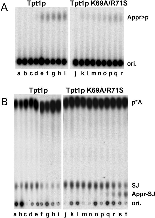
Tpt1p is an essential protein responsible for the 2'-phosphotransferase step of tRNA splicing in Saccharomyces cerevisiae, in which the splice junction 2'-phosphate of ligated tRNA is transferred to NAD to form mature tRNA and ADP-ribose 1''-2'' cyclic phosphate. We showed previously that Tpt1p is a member of a family of functional 2'-phosphotransferases found in eukaryotes, eubacteria, and archaea, that the Escherichia coli protein (KptA) is highly specific for 2'-phosphorylated RNAs despite the lack of obvious natural substrates, and that KptA acts on a trinucleotide substrate through an intermediate in which RNA is ADP-ribosylated at the 2'-phosphate. This mechanism is similar to a proposed mechanism of NAD-dependent histone deacetylases. We present evidence here that this mechanism is conserved in S. cerevisiae, and we identify residues important for the second step of the reaction, during which the intermediate is resolved into products. We examined 21 Tpt1 protein variants mutated in conserved residues or blocks of residues and show that one of them, Tpt1 K69A/R71S protein, accumulates large amounts of intermediate with trinucleotide substrate due to a very slow second step. This intermediate can be trapped on beads when formed with biotin-NAD. We also show that Tpt1 K69A/R71S protein forms an intermediate with the natural ligated tRNA substrate and demonstrate that, as expected, this mutation is lethal in yeast. The high degree of conservation of these residues suggests that the entire Tpt1p family is involved in a similar two-step chemical reaction.


Figures

FIGURE 1.
FIGURE 1.
A schematic of the second step of the 2′-phosphotrans-ferase reaction. In the first step the 2′-phosphate of the RNA substrates attacks NAD, generating ADP-ribosylated phosphorylated RNA, and releasing nicotinamide (Spinelli et al. 1999). In the second step, a presumed transesterification as shown generates ADP-ribose 1″-2″-cyclic phosphate and dephosphorylated RNA product.
FIGURE 2.
FIGURE 2.
Amino acid alignment of 2′-phosphotransferases. The amino acid alignment shown contains 2′-phosphotransferase orthologs from M. musculus, Schizosaccharomyces pombe, Arabidoposis thaliana, Escherichia coli, A. fulgidis, and Saccharomyces cerevisiae. The sequences resulted from a BLAST search of the Saccharomyces cerevisiae sequence; the alignment was done using clustalW (http://searchlauncher.bcm.tmc.edu/multi-align.html) and box-shade (http://www.ch.embnet.org/software/boxform.html). The positions of K69 and R71 are shown with arrows.
FIGURE 3.
FIGURE 3.
Titration of Tpt1 K69A/R71S and Tpt1 protein with tri-nucleotide substrate. Tpt1 K69A/R71S protein and Tpt1 protein were assayed for 2′-phosphotransferase activity as described in Materials and Methods, in 10 μL reaction mixtures containing 1 fmole p*ApAppA, the indicated concentration of NAD, and enzyme, for 30 min at 30°C, and products were separated by thin-layer chromatography as described in Materials and Methods. a, no protein; bg, hm, fivefold serial titrations of Tpt1 K69A/R71S protein, starting with 4.3 μM protein, at 0.25 mM NAD (bg) and at 10 mM NAD (hm); nr, sw, fivefold serial titrations of Tpt1 protein, starting with 280 nM protein at 0.25 mM NAD (nr) and at 10 mM NAD (sw).
FIGURE 4.
FIGURE 4.
Time course of reaction with Tpt1 protein or Tpt1p K69A/R71S and trinucleotide substrate. Tpt1 K69A/R71S protein (2.2 μM) and Tpt1 protein (0.14 nM) were assayed for 2′-phosphotransferase activity as described in Materials and Methods in 50 μL reaction mixtures containing 2 μM p*ApAppA substrate and 10 mM NAD, and 3 μL aliquots were sampled at different times, spotted to PEI cellulose plates, and resolved in buffer containing 2M sodium formate (pH 3.5). (A) Time course of Tpt1 protein activity. (Lane a) No protein, 0 min; (lanes bm) products formed with Tpt1 protein, at 0, 1, 2, 5, 10, 15, 20, 30, 45, 60, 75, 90 min. (B) Time course of Tpt1 K69A/R71S protein activity. (Lanes al) Products formed with Tpt1 K69A/R71S protein, at 0, 1, 2, 5, 10, 15, 20, 30, 45, 60, 75, 90 min; (lane m) no protein, 90 min. (C,D) Quantification of time course of reactions of Tpt1 protein and Tpt1 K69A/R71S protein with trinucleotide substrate. Products, intermediate, and substrate were quantified by PhosphorImager and plotted using SigmaPlot.
FIGURE 5.
FIGURE 5.
The intermediate formed with Tpt1K69A/R71S protein contains NAD. (A) Intermediate is formed with labeled NAD. Reaction mixtures containing 5 nM pApAppA substrate and 0.5 nM Ap*pN, as well as the indicated amount of unlabeled NAD, were incubated with no protein (lanes a,e), 0.7 μM Tpt1 protein (lanes bd), or 2.2 μM Tpt1 K69A/R71S protein (lanes fh). (B) Intermediate is formed with biotin-NAD. Reaction mixtures containing 1 nM p*ApAppA and 75 μM NAD or biotin-NAD, as indicated, were incubated with protein, and products were resolved on thin-layer plates as described. (Lanes ac) Buffer; (lanes dh) Tpt1 K69A/R71S protein at 4.3 μM (lanes d,f,h) or 2.2 μM (lanes e,g); (lanes ik) 0.7 μM Tpt1 protein.
FIGURE 6.
FIGURE 6.
Intermediate formed with biotin-NAD can be resolved on streptavidin beads. Reaction mixtures containing 1 nM p*ApAppA substrate, 12 μM Tpt1 K69A/R71S protein, and 75 μM NAD (lanes bd) or biotin NAD (lanes el) were incubated for 40 min and then incubated with streptavidin beads as described in Materials and Methods. (Lane a) No protein; (lanes b,d) reaction mixture; (lanes c,f) first wash after streptavidin beads; (lanes d,g) second wash after streptavidin beads; (lane h) washed beads extracted with phenol; (lane j) reaction mixture (from lane e) extracted with phenol; (lane i) mixture of material from lanes h and j, cospotted on thin-layer plate; (lane k) washed beads treated with Tpt1p; (lane l) reaction mixture (from lane e) treated with Tpt1p. R, reaction mixture; w1, w2 eluant from washes of beads; B, beads after washes; cs, cospotting.
FIGURE 7.
FIGURE 7.
Detection of the reaction intermediate with Tpt1 K69A/R71S protein and ligated tRNA substrate. (A) NAD dependence of Appr>p formation from ligated tRNA. 50 pM Tpt1p (lanes ai) or 2.2 μM Tpt1 K69A/R71S protein (lanes jr) was incubated with 0.15 nM ligated tRNA and increasing concentrations of NAD, and products were resolved on thin-layer plates as described above. (Lanes a,j) No NAD; (lanes bi,kr) 2 nM, 20 nM, 200 nM, 2 μM, 20 μM, 200 μM, 2 mM, and 20 mM NAD. (B) NAD dependence of intermediate formation from ligated tRNA. Samples from A above were treated with P1 nuclease, and products were resolved on thin-layer plates. SJ, splice junction; Appr-SJ, ADP-ribosylated splice junction.

References

    1. Abelson, J., Trotta, C.R., and Li, H. 1998. tRNA splicing. J. Biol. Chem. 273: 12685–12688. - PubMed
    1. Barnard, E.A. 1969. Ribonucleases. Annu. Rev. BioChem. 38: 677–732. - PubMed
    1. Belford, H.G., Westaway, S.K., Abelson, J., and Greer, C.L. 1993. Multiple nucleotide cofactor use by yeast ligase in tRNA splicing. Evidence for independent ATP- and GTP-binding sites. J. Biol. Chem. 268: 2444–2450. - PubMed
    1. Bell, C.E. and Eisenberg, D. 1996. Crystal structure of diphtheria toxin bound to nicotinamide adenine dinucleotide. Biochemistry 35: 1137–1149. - PubMed
    1. Bradford, M.M. 1976. A rapid and sensitive method for the quantitation of microgram quantities of protein utilizing the principle of protein-dye binding. Anal. BioChem. 72: 248–254. - PubMed

Publication types

MeSH terms

LinkOut - more resources