. 2005 Jan 4;102(1):128-33.
doi: 10.1073/pnas.0403198102. Epub 2004 Dec 20.

Microvascular patterning is controlled by fine-tuning the Akt signal

Affiliations

Microvascular patterning is controlled by fine-tuning the Akt signal

Jing Fang Sun et al. Proc Natl Acad Sci U S A. .

Abstract

We investigated the functions of Akt during vascular development and remodeling by using an inducible endothelial cell-specific driver of the dominant-active myrAkt. We found that sustained signaling in response to overexpression of myrAkt led to embryonic lethality, edema, and vascular malformations. In addition to the morphological malformations, the vascular phenotype was consistent with a failure in remodeling, such that the normal patterning and vessel hierarchy was disturbed. Examination of the well studied retinal vasculature during the remodeling phases revealed that transient expression of myrAkt was capable of altering the normal response to oxygen-induced remodeling without causing vascular malformations. These findings suggest that physiological levels of Akt signaling modulated microvascular remodeling and support the hypothesis that, although Akt may be required for vascular growth and homeostasis, appropriate down-regulation is also an essential aspect of normal vascular patterning.


Figures

Fig. 1.
Fig. 1.
Survival functions in retinal remodeling. (a) Microvascular remodeling during normal retinal development is shown after Lectin BS-1 (Sigma) staining to highlight the lack of vessel definition before remodeling in a P3 mouse, followed by the formation of a CFZ that defines arterial vessels by P7, and the further extension of capillary reduction from the arterial vessel toward the vein by P13 in the region closest to the optic disk, which is at the left edge of all of these images. (b) FACS histograms of DNA content for negative (0% FBS) and positive (10% FBS) control are shown on left, and results listed for all factors tested are shown on right. (c) The acceleration of capillary reduction is achieved by hyperoxia exposure. The CFZ doubles in size from 0 to 6 h, and by 24 h total clearing of microvessels from artery to vein is observed in a circular region surrounding the optic disk. The image for 24 h is half the magnification of the rest to show distance of clearing. (d) This exaggerated formation of a CFZ is coincident with decreased Akt phosphorylation as marked by Western blot for pAkt (BD Pharmingen). Total Akt (Santa Cruz Biotechnology) and actin (Sigma) serve as protein loading controls. a, arterial vessel; v, vein; *, CFZ.
Fig. 2.
Fig. 2.
Sustained endothelial cell-specific expression of myrAkt driven by VE-cadherin promoter-tTA transgenic mice induces microvascular malformations in the mature retina. VE-cadherin:tTA mice crossed to TET:lacZ mice shows β-galactosidase expression in embryonic blood vessels (E14.5) (a), retinal blood vessels (b), and costaining of β-galactosidase with anti-VEGFR-2 (c). (d) Western blot analysis of retinas from adults and neonates show transgene expression by using hemagglutinin (HA) tag (Roche Diagnostics) and increased levels of phosphorylated Akt (BD Pharmingen). (e) Sustained expression of myrAkt causes malformations reminiscent of microaneurysms in the retina. (f) Red Lectin-TRITC staining. Retinal vessel diameter from double transgenic pups (seven animals) is significantly larger than vessel diameter from control pups (four animals) after adjusting for litter effects by stratified Wilcoxon–Mann–Whitney test, P < 0.0001.
Fig. 3.
Fig. 3.
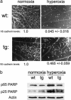
Transient expression of myr-Akt impairs oxygen-induced reductions in microvessel density and apoptosis during remodeling. (a) Whole-mount immunohistochemistry of retinas using BS-I Lectin-TRITC on control and double transgenic P7 neonatal mice before (normoxia) and after (hyperoxia) show the block in exaggerated capillary clearing coincident with Akt induction. VE-cadherin mRNA levels were quantified by real-time RT-PCR and shown below each panel relative to normoxia control. (b) Western blot analysis of PARP cleavage was used to quantify apoptosis in retinas after hyperoxia with and without transgenic myrAkt expression. The PARP antibody (Biosource) recognizes the p25 and p85 cleavage products. wt, wild type; tg, double transgenic.
Fig. 4.
Fig. 4.
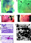
Inducible myr-Akt blocks normal remodeling in embryonic skin. Tie-2:lacZ transgenic mice were used to visualize blood vessels in embryo at E12.5 (a) and E14.5 (b) to show remodeling of the immature capillary network into arbor-like vessels. Compared to the normal embryonic vasculature (c), in double transgenic VE-cadherin:tTA;TET:myrAkt embryos, this remodeling is abnormal and an arbor-like hierarchy does not form (d). (e) Histology of wild-type embryonic skin as a control. (f) Double transgenic skin shows edema in s.c. adipose tissue, disruption of the skeletal muscle layer (highlighted with an arrow), and congested and large vessels in the dermis. Note the increased size of the connective tissue layer, as indicated with a bar on the right. (d, dermis; c, connective tissue). Images are the same magnification. (g) Electron microscopy of double transgenic embryos reveals frank disruptions of the vessel wall and erythrocyte leakage without evidence of endothelial cell necrosis or apoptosis (e, endothelial cell; p, pericyte).

References

    1. Dimmeler, S. & Zeiher, A. M. (2000) Circ. Res. 87, 434-439. - PubMed
    1. Liu, W., Ahmad, S. A., Reinmuth, N., Shaheen, R. M., Jung, Y. D., Fan, F. & Ellis, L. M. (2000) Apoptosis 5, 323-328. - PubMed
    1. Stone, J., Itin, A., Alon, T., Pe'er, J., Gnessin, H., Chan-Ling, T. & Keshet, E. (1995) J. Neurosci. 15, 4738-4747. - PMC - PubMed
    1. Provis, J. M., Leech, J., Diaz, C. M., Penfold, P. L., Stone, J. & Keshet, E. (1997) Exp. Eye Res. 65, 555-568. - PubMed
    1. Pe'er, J., Shweiki, D., Itin, A., Hemo, I., Gnessin, H. & Keshet, E. (1995) Lab. Invest. 72, 638-645. - PubMed

Publication types

MeSH terms

Substances

LinkOut - more resources