. 2005 Jan;79(2):732-44.
doi: 10.1128/JVI.79.2.732-744.2005.

Involvement of influenza virus PA subunit in assembly of functional RNA polymerase complexes

Affiliations

Involvement of influenza virus PA subunit in assembly of functional RNA polymerase complexes

Atsushi Kawaguchi et al. J Virol. 2005 Jan.

Abstract

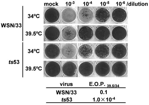
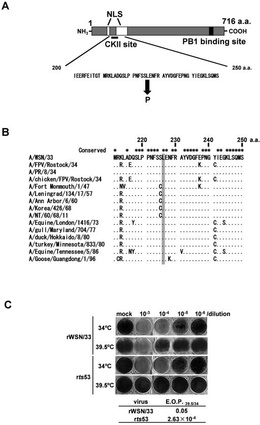
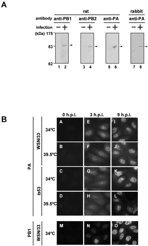
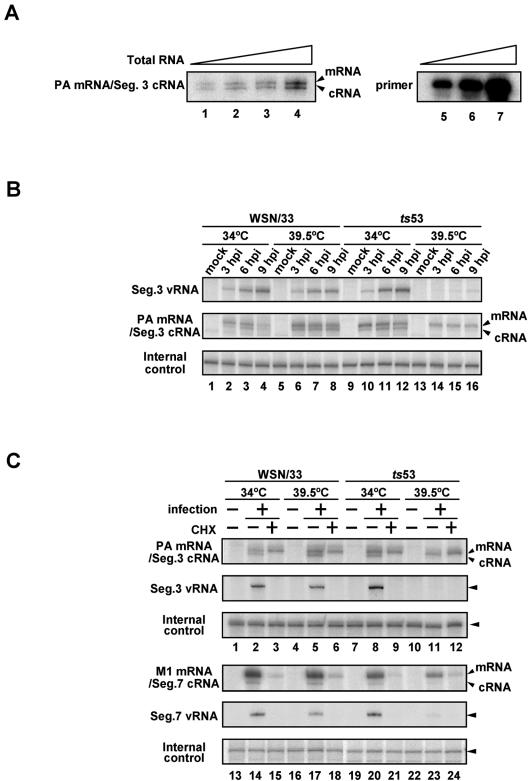
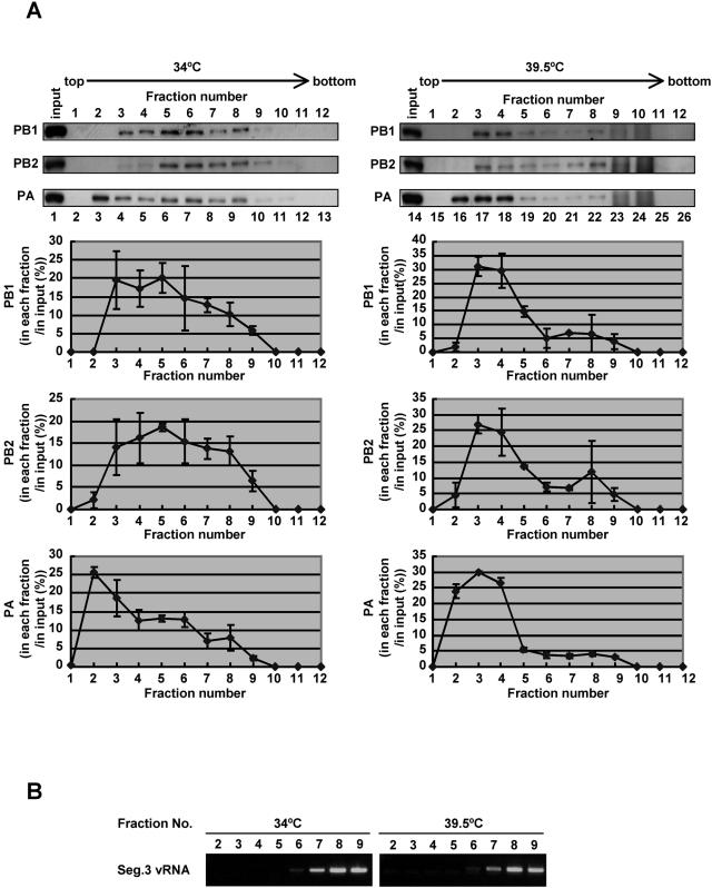
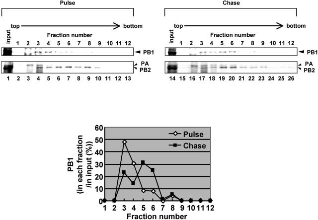
The RNA-dependent RNA polymerase of influenza virus consists of three subunits, PB1, PB2, and PA, and synthesizes three kinds of viral RNAs, vRNA, cRNA, and mRNA. PB1 is a catalytic subunit; PB2 recognizes the cap structure for generation of the primer for transcription; and PA is thought to be involved in viral RNA replication. However, the process of polymerase complex assembly and the exact nature of polymerase complexes involved in synthesis of the three different RNA species are not yet clear. ts53 virus is a temperature-sensitive (ts) mutant derived from A/WSN/33 (A. Sugiura, M. Ueda, K. Tobita, and C. Enomoto, Virology 65:363-373, 1975). We confirmed that the mRNA synthesis level of ts53 remains unaffected at the nonpermissive temperature, whereas vRNA synthesis is largely reduced. Sequencing of the gene encoding ts53 PA and recombinant virus rescue experiments revealed that an amino acid change from Leu to Pro at amino acid position 226 is causative of temperature sensitivity. By glycerol density gradient analyses of nuclear extracts prepared from wild-type virus-infected cells, we found that polymerase proteins sediment in three fractions: one (H fraction) consists of RNP complexes, another (M fraction) contains active polymerases but not viral RNA, and the other (L fraction) contains inactive forms of polymerases. Pulse-chase experiments showed that polymerases in the L fraction are converted to those in the M fraction. In ts53-infected cells, polymerases accumulated in the L fraction. These results strongly suggest that PA is involved in the assembly of functional viral RNA polymerase complexes from their inactive intermediates.


Figures

FIG. 1.
FIG. 1.
Temperature sensitivity of ts53 virus in plaque formation. Plaque assays were carried out at 34°C (permissive temperature) and 39.5°C (nonpermissive temperature) with diluted (103 to 106 times) wild-type WSN/33 virus and ts53 virus. The efficiency of plaque formation of WSN/33 virus at 39.5°C relative to that at 34°C (EOP39.5/34) was 0.1, whereas the EOP39.5/34 of ts53 virus was 1.0 × 10⁻⁴.
FIG. 2.
FIG. 2.
Mutation point in the PA gene of ts53 virus. (A) A single point mutation is located near nuclear localization signals (NLS) and putative phosphorylation sites. This mutation causes an amino acid change from Leu to Pro at amino acid position 226. The PB1 binding site is also shown (28). (B) The conservation of Leu at amino acid position 226 among several influenza A viruses. The mutated amino acid in the PA of ts53 is indicated by a gray box. This amino acid is highly conserved among several influenza A viruses. (C) Responsibility of the mutation at amino acid position 226 for the temperature sensitivity of ts53 virus. A recombinant virus was generated by reverse genetics that contains the ts53 PA gene and seven other genes derived from wild-type virus. Plaque assays were carried out at 34 or 39.5°C. The EOP39.5/34 of recombinant WSN/33 virus was 0.05, whereas the EOP39.5/34 of recombinant ts53 virus was 2.63 × 10⁻⁴. a.a., amino acid.
FIG. 3.
FIG. 3.
Intracellular localization of ts53 PA. (A) Generation of rat polyclonal antibodies against PB1, PB2, and PA and a rabbit polyclonal antibody against PA. Details of the protocol for preparation of the antibodies are described in Materials and Methods. Lysates prepared from WSN/33 virus-infected MDCK cells (lanes 2, 4, 6, and 8) or mock-infected MDCK cells (lanes 1, 3, 5, and 7) were subjected to Western blotting using each antibody. Proteins corresponding to antigens are indicated by arrowheads. (B) Intracellular localization of ts53 PA. MDCK cells were infected with WSN/33 or ts53 virus at 34 or 39.5°C at an MOI of 10. At 3 (panels E to H and N) and 9 (panels I to L and O) h postinfection (h.p.i.), cells were fixed with 4% paraformaldehyde and indirect immunofluorescence assays were carried out using rat anti-PA antibody (panels A to L) and rat anti-PB1 antibody (panels M to O) as described in Materials and Methods. Mock-infected cells were also examined (panels A to D and M).
FIG. 4.
FIG. 4.
Temperature sensitivity of viral RNA synthesis in ts53 virus-infected cells. Primer extension assays were performed with 4 μg of total RNAs and vRNA- or mRNA/cRNA-specific DNA primers labeled at their 5′ end with [γ-32P]ATP. Reverse-transcribed products were analyzed on a 5% polyacrylamide gels containing 7 M urea in TBE buffer and were detected by autoradiography. (A) Dose response in the primer extension assay. Total RNAs were purified from WSN/33 virus-infected MDCK cells at 6 h postinfection (hpi). Primer extension assays were performed with 1 (lane 1), 2 (lane 2), 4 (lane 3), and 8 μg (lane 4) of total RNA and a primer specific for segment (Seg.) 3 mRNA/cRNA as described in Materials and Methods. To show that the amount of the primer was in excess in the primer extension assay, 1/400 (lane 5), 1/200 (lane 6), and 1/100 (lane 7) of the input primers were also subjected to electrophoresis. (B) Synthesis of viral RNAs in ts53 virus-infected cells. MDCK cells were infectedwith WSN/33 or ts53 virus at an MOI of 10. At 3 (lanes 2, 6, 10, and 14), 6 (lanes 3, 7, 11, and 15), and 9 (lanes 4, 8, 12, and 16) h postinfection at 34°C (lanes 1 to 4 and 9 to 12) or 39.5°C (lanes 5 to 8 and 13 to 16), cells were collected and total RNAs were isolated. Primer extension assays were performed with primers specific for segment 3 vRNA or mRNA/cRNA. Total RNAs prepared from uninfected cells were also analyzed (lanes 1, 5, 9, and 13). As an internal control, luciferase mRNA (10 ng) was added to each sample, and the sample was then subjected to primer extension assays. (C) Viral mRNA synthesis in the presence of a protein synthesis inhibitor. MDCK cells were infected with WSN/33 or ts53 virus at an MOI of 10 in the presence or absence of 100 μg of cycloheximide (CHX)/ml. At 6 h postinfection in the presence (lanes 3, 6, 9, 12, 15, 18, 21, and 24) or absence (lanes 2, 5, 8, 11, 14, 17, 20, and 23) of CHX at 34 or 39.5°C, total RNAs were isolated. Primer extension assays were performed with primers specific for vRNA and mRNA/cRNA of segment 3 and segment 7. Total RNAs prepared from uninfected cells without CHX were also analyzed (lanes 1, 4, 7, 10, 13, 16, 19, and 22). luciferase mRNA was used as an internal control for the whole procedure.
FIG. 5.
FIG. 5.
Fractionation of polymerase complexes in ts53-infected cells. MDCK cells were infected with ts53 virus at an MOI of 10 at 34 or 39.5°C for 6 h. Cells were collected and permeabilized in 100 μg of digitonin/ml. Soluble nuclear lysates prepared from permeabilized cells were loaded onto a 30 to 60% glycerol density gradient and centrifuged at 4°C in an SW28 rotor at 45,000 rpm for 5 h. Fractions (1 to 12) were recovered from the top of the gradient. (A) Western blotting analyses. For the upper panel, an aliquot of each fraction was separated in a SDS-7.5% PAGE and subjected to Western blotting using rabbit anti-PB1, anti-PB2, and anti-PA antibodies. Lanes 2 to 13 and lanes 15 to 25 correspond to glycerol gradient fractions of lysates prepared from cells infected at 34 and 39.5°C, respectively. Lysates loaded onto the glycerol density gradient are alsoshown in lanes 1 and 14. The lower panel shows results of the upper panel quantitatively analyzed by the NIH image analyzing system for determination of the amounts of PB1, PB2, and PA in each fraction. The ratio of the amount of PB1, PB2, or PA in each fraction relative to that in the input (the total amount of each polymerase protein) is indicated. (B) RNA analysis. RNAs were purified from glycerol density gradient fractions (fractions 2 to 9), and the segment (Seg.) 3 vRNA was semiquantitatively analyzed by reverse transcription-PCR. cDNA was synthesized by reverse transcriptase with PA-1356 as the primer. Single-stranded cDNA was then PCR amplified with two specific primers, PA-1683 primer (5′-GGCACTTCTTAGAAGCATATCTC-3′), corresponding to the PA coding region between nucleotide sequence positions 1661 and 1683, and PA-1356 primer (PCR cycles consisted of 20 cycles for samples obtained from 34°C assays and 25 cycles for those from 39.5°C assays). Amplified double-stranded DNAs were subjected to a 1% agarose gel electrophoresis and were visualized by ethidium bromide.
FIG. 6.
FIG. 6.
Conversion of polymerase complexes. MDCK cells were infected with ts53 virus at an MOI of 10 at 34°C for 4.5 h. Cells were washed and incubated in methionine-free medium for 30 min. Infected cells were then pulse labeled at 34°C for 15 min in the presence of 10 μCi of [35S]methionine and [35S]cysteine mixture/ml. After being washed with MEM, cells were further incubated at 34°C for 15 (pulse) or 60 min (chase) in MEM containing excess amounts of nonlabeled methionine and cysteine. Lysates prepared from metabolically labeled cells were subjected to the glycerol density gradient centrifugation assay as described in the legend to Fig. 5. Upper panel, fractionation of pulse-labeled and pulse-chased polymerases. An aliquot of each fraction was separated in SDS-6% PAGE and then was visualized by autoradiography. PB1, PB2, and PA are indicated by arrowheads. We confirmed that these bands correspond to PB1, PB2, and PA by immunoprecipitation assay using antibodies specific for each polymerase subunit (data not shown). (Lower panel) To determine the amount of PB1 in each fraction, autoradiograms were quantitatively analyzed by the NIH image analyzing system, and the amount of PB1 in each fraction is shown as that relative to the total amount of PB1.
FIG. 7.
FIG. 7.
Properties of polymerase complexes. (A) Recognition specificity of PA by the rabbit anti-PA antibody. vRNP obtained from purified virions was boiled at 100°C for 3 min with 2% SDS (lanes 1 to 3). The solution was then diluted with the IP buffer so that the final concentration of SDS was set at 0.04%. SDS-disrupted vRNP (lanes 1 to 3) and undisrupted vRNP (lanes 4 to 6) were subjected to immunoprecipitation with (lanes 1, 2, 4, and 5) or without (lanes 3 and 6) the rabbit anti-PA antibody as described in Materials and Methods. Immunoprecipitated proteins were separated in SDS-7.5% PAGE and were detected by Western blotting with the rat anti-PA antibody. Lane 7 shows the input vRNP (80 ng of PA). For lanes 1 and 4 and lanes 2, 3, 5, and 6, aliquots containing 400 and 800 ng of PA were used, respectively. (B) Immunoprecipitation of fractions obtained from the glycerol density gradient centrifugation. The fractions (50 μl) corresponding to those in Fig. 5A (lanes 3 to 10 and 16 to 23) were subjected to immunoprecipitation with the rabbit anti-PA antibody (lanes 1 to 8 and 9 to 16, respectively). Immunoprecipitated proteins were separated in SDS-7.5% PAGE and were detected by Western blotting by rat anti-PB1 and anti-PA antibodies. (C) In vitro RNA synthesis using fractions recovered from glycerol density gradient centrifugation. In vitro RNA synthesis was carried out with fraction 3 (lanes 1 and 2), fraction 5 (lanes 3 and 4), and vRNP (lanes 5 and 6; positive controls) prepared from purified virion as enzyme sources, essentially as described previously (, , ; for details, see the text). [α-32P]GTP-labeled products were separated on a 10% polyacrylamide gel containing 8 M urea and were visualized by autoradiography. Aliquots of fractions 3 and 5 containing 1.25 (lanes 1 and 3) and 5 ng (lanes 2 and 4) of PB1 and vRNP complexes containing 0.25 and 1 ng of PB1 (lanes 5 and 6, respectively) were used.

References

    1. Akkina, R. K., T. M. Chambers, D. R. Londo, and D. P. Nayak. 1987. Intracellular localization of the viral polymerase proteins in cells infected with influenza virus and cells expressing PB1 protein from cloned cDNA. J. Virol. 61:2217-2224. - PMC - PubMed
    1. Belz, G. T., W. Xie, J. D. Altman, and P. C. Doherty. 2000. A previously unrecognized H-2D(b)-restricted peptide prominent in the primary influenza A virus-specific CD8(+) T-cell response is much less apparent following secondary challenge. J. Virol. 74:3486-3493. - PMC - PubMed
    1. Biswas, S. K., and D. P. Nayak. 1994. Mutational analysis of the conserved motifs of influenza A virus polymerase basic protein 1. J. Virol. 68:1819-1826. - PMC - PubMed
    1. Braam, J., I. Ulmanen, and R. M. Krug. 1983. Molecular model of a eucaryotic transcription complex: functions and movements of influenza P proteins during capped RNA-primed transcription. Cell 34:609-618. - PubMed
    1. Fodor, E., M. Crow, L. J. Mingay, T. Deng, J. Sharps, P. Fechter, and G. G. Brownlee. 2002. A single amino acid mutation in the PA subunit of the influenza virus RNA polymerase inhibits endonucleolytic cleavage of capped RNAs. J. Virol. 76:8989-9001. - PMC - PubMed

Publication types

LinkOut - more resources