. 2005 Jan;79(2):841-52.
doi: 10.1128/JVI.79.2.841-852.2005.

Epstein-Barr virus gp42 is posttranslationally modified to produce soluble gp42 that mediates HLA class II immune evasion

Affiliations

Epstein-Barr virus gp42 is posttranslationally modified to produce soluble gp42 that mediates HLA class II immune evasion

Maaike E Ressing et al. J Virol. 2005 Jan.

Abstract

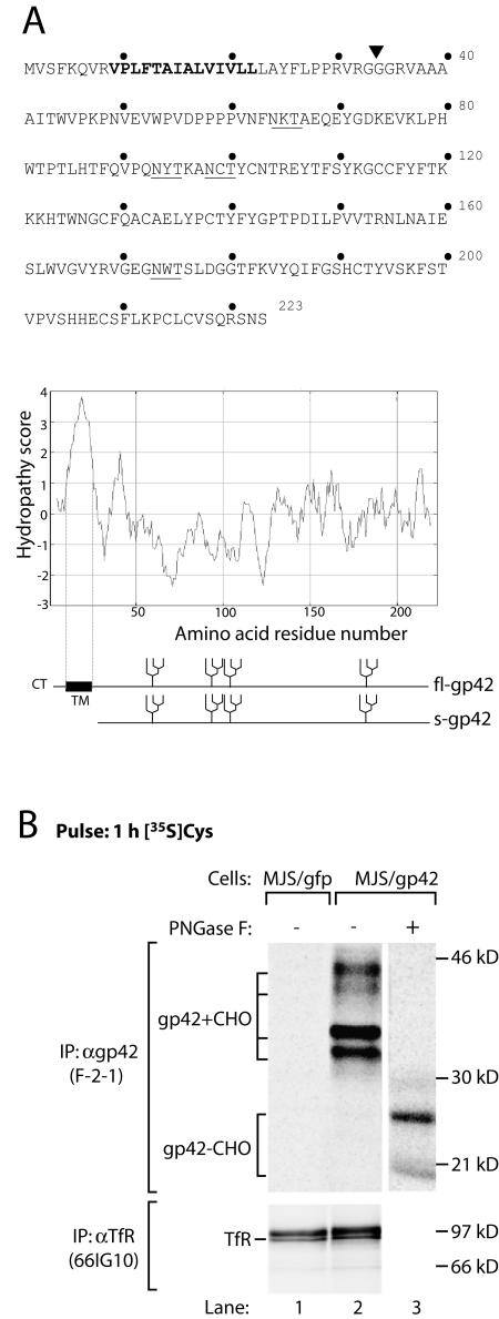
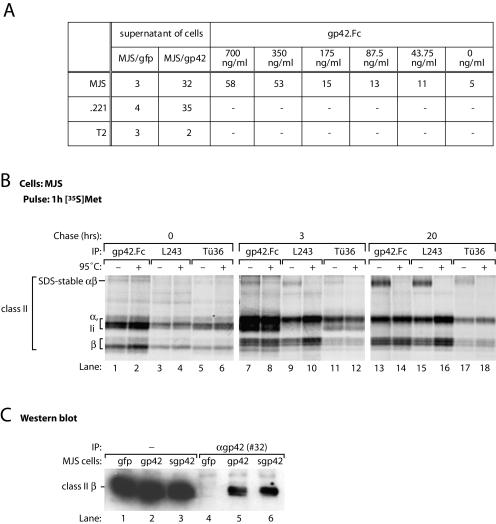
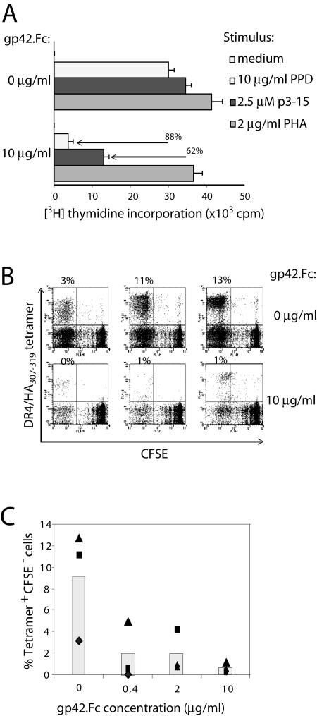
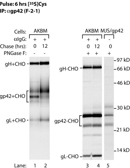
Epstein-Barr virus (EBV) resides as a persistent infection in human leukocyte antigen (HLA) class II+ B lymphocytes and is associated with a number of malignancies. The EBV lytic-phase protein gp42 serves at least two functions: gp42 acts as the coreceptor for viral entry into B cells and hampers T-cell recognition via HLA class II molecules through steric hindrance of T-cell receptor-class II-peptide interactions. Here, we show that gp42 associates with class II molecules at their various stages of maturation, including immature alphabetaIi heterotrimers and mature alphabeta-peptide complexes. When analyzing the biosynthesis and maturation of gp42 in cells stably expressing the viral protein, we found that gp42 occurs in two forms: a full-length type II membrane protein and a truncated soluble form. Soluble gp42 is generated by proteolytic cleavage in the endoplasmic reticulum and is secreted. Soluble gp42 is sufficient to inhibit HLA class II-restricted antigen presentation to T cells. In an almost pure population of Burkitt's lymphoma cells in the EBV lytic cycle, both transmembrane and soluble forms of gp42 are detected. These results imply that soluble gp42 is generated during EBV lytic infection and could contribute to undetected virus production by mediating evasion from T-cell immunity.


Figures

FIG. 1.
FIG. 1.
EBV gp42 occurs in two forms. (A) Amino acid sequence (single-letter code) and Kyte-Doolittle hydropathy plot of EBV gp42. Bold type highlights the putative transmembrane domain. The N-linked glycosylation sites are underlined. A signal sequence cleavage site is predicted between residues 33 and 34 (inverted triangle); fl-gp42 and the truncated protein potentially generated upon cleavage (s-gp42) are depicted. CT, cytoplasmic tail; TM, transmembrane domain. (B) MJS/gp42 cells (lanes 2 and 3) and control MJS/gfp cells (lane 1) were metabolically labeled for 1 h with [35S]Cys (gp42) or with[35S]Met (TfR). After lysis of the cells, MAb F-2-1 was used to isolate EBV gp42 molecules. Portions of the immunoprecipitates were treated with PNGase F to remove N-linked glycans. Glycosylated (gp42+CHO) and nonglycosylated (gp42−CHO) gp42 polypeptides are indicated. As a control protein, TfR was precipitated with MAb 66IG10. Samples were analyzed by reducing SDS-12% PAGE. IP, immunoprecipitation; α, anti-.
FIG. 2.
FIG. 2.
A truncated form of EBV gp42 is secreted over time. (A) MJS/gp42 cells were labeled for 30 min with [35S]Cys and chased for the times indicated. EBV gp42 molecules were recovered from cell lysates (lanes 1 to 7) and from supernatants (lanes 8 to 13) with MAb F-2-1. To reveal the protein backbones, portions of the immunoprecipitates were treated with PNGase F (lanes 7 and 13). (B) Digestions with endo H were performed to examine protein transport beyond the ER and cis-Golgi compartments; endo H-sensitive (endo HS) and endo H-resistant (endo HR) gp42 forms are indicated. All samples were analyzed by SDS-12% PAGE. Untreated F-2-1 precipitates were boiled in nonreducing sample buffer, whereas PNGase F- and endo H-digested samples were denatured under reducing conditions. (C) EBV gp42 was analyzed after in vitro translation of gp42-4Met mRNA in the presence (lanes 2 to 4) or in the absence (lane 1) of microsomes. Portions of the samples were digested with saturating (lane 3) or suboptimal (lane 4) amounts of PNGase F. fl-gp42 molecules bearing one to four N-linked glycans are indicated (+1 through +4); fl-gp42+CHO and s-gp42+CHO overlap such that, for instance, fl-gp42 plus one N-linked glycan runs with a mobility comparable to that of s-gp42 plus two N-linked glycans. See the legend to Fig. 1 for definitions of abbreviations.
FIG. 3.
FIG. 3.
s-gp42 is capable of binding to both immature and mature HLA-DR molecules. (A) Culture supernatants of MJS/gp42 cells and control MJS/gfp cells were incubated with HLA class II+ MJS and .221 cells or with HLA class II T2 cells; for comparison, the binding of gp42.Fc was included. Cell-associated gp42 was detected with antiserum #32 and analyzed by flow cytometry. Mean fluorescence values are depicted; −, not tested. (B) MJS cells were metabolically labeled with [35S]Met for 1 h and chased for 3 or 20 h before lysis. Immunoprecipitation (IP) was performed with s-gp42.Fc or HLA-DR-specific MAbs L243 and Tü36. Immune complexes were incubated in nonreducing sample buffer for 3 h at 37°C (−) or for 5 min at 95°C (+) prior to separation by SDS-12% PAGE. (C) Total lysates of MJS/gp42 cells (lanes 2 and 5), MJS/Kbss-gp42 cells (lanes 3 and 6), and control MJS/gfp cells (lanes 1 and 4) were used directly (lanes 1 to 3) or were subjected to immunoprecipitation with rabbit antiserum #32 against gp42 (lanes 4 to 6). Total lysates and immune complexes were boiled in nonreducing sample buffer, separated by SDS-12% PAGE, and blotted onto polyvinylidene difluoride membranes. Western blots were stained with a MAb specific for HLA-DR β chains (HB10A) and visualized by enhanced chemiluminescence.
FIG. 4.
FIG. 4.
EBV gp42 associates with HLA-DR molecules intracellularly shortly after biosynthesis. (A) MJS/gp42 cells were metabolically labeled with [35S]Cys (lanes 1 to 6) or with [35S]Met (lanes 7 to 18) for 30 min and chased for the times indicated. For immunoprecipitation (IP), rabbit antiserum #32 against gp42 and MAb Tü36 against HLA-DR were used. (B) Half of the immunoprecipitates were treated with PNGase F to remove N-linked glycans. Immune complexes were denatured in nonreducing (A) or reducing (B) sample buffer and analyzed by SDS-12% PAGE.
FIG. 5.
FIG. 5.
Treatment with ConB prevents the degradation of EBV gp42 and stabilizes gp42-HLA-DR complexes. MJS/gp42 cells (A and B, upper panels) and control MJS/gfp cells (B, lower panels) were mock treated (−) or treated (+) with ConB during starvation, labeling, and chase to prevent endolysosomal proteolysis. Pulse-labeling was performed for 1 h with [35S]Cys (A) or [35S]Met (B); chase times were 4 and 20 h. EBV gp42 molecules were isolated from cell lysates (lanes 1 to 12) and from supernatants (lanes 13 to 18) with antiserum #32 (A and B, lanes 1 to 6) or MAb F-2-1, which is specific for free gp42 (A, lanes 7 to 18); HLA-DR complexes were precipitated with MAb Tü36 (B, lanes 7 to 12). All samples were analyzed by nonreducing SDS-12% PAGE. IP, immunoprecipitation.
FIG. 6.
FIG. 6.
s-gp42 is sufficient to inhibit antigen-specific HLA-DR-restricted T-cell recognition. (A) Effects of gp42.Fc on activation of M. tuberculosis-specific Rp15.1.1 T cells were examined upon coculturing with HLA-DR3+ PBMC in the absence or in the presence of PPD as a source of hsp65, the related p3-15 peptide, or the mitogen PHA. [3H]Thymidine incorporation is shown with error bars for triplicate samples. (B) TCR-HLA-DR-peptide interactions in the presence of various concentrations of gp42.Fc were evaluated with PBMC that were obtained from an HLA-DR4+ donor, labeled with CFSE, and stimulated in vitro with HA307-319 peptides. After 2 weeks, responding T cells were incubated with specific allophycocyanine-conjugated tetramers and were stained with PE-conjugated MAbs to CD4. To study the influence of gp42.Fc on recall responses induced by TCR-HLA-DR-peptide, we used the same approach, except that gp42.Fc was provided during the 2-week in vitro stimulation. Responding T cells were incubated with HLA-DR4/HA307-319 tetramers and stained for CD4. Each flow cytometry dot plot represents approximately 30,000 CD4+ PI cells; percent values represent the percentages of tetramer-positive CFSE cells among the total CD4+ PI T cells (upper leftquadrant). (C) The percentages of tetramer-positive CFSE T cells (among the CD4+ PI cells) in the experiment shown in panel B are plotted against the concentration of gp42.Fc. The symbols (▴, ▪, and ♦) represent the three wells of the triplicates.
FIG. 7.
FIG. 7.
During EBV lytic infection of B cells, both forms of gp42 are generated. AKBM cells were cultured with anti-IgG antibody to induce EBV reactivation. At 20 h postinduction, AKBM cells in the lytic cycle were positively selected for the expression of the inducible rat CD2-GFP reporter protein, pulse-labeled for 6 h with [35S]Cys, and chased for up to 12 h prior to NP-40 cell lysis. SDS-12% PAGE analysis is shown for proteins immunoprecipitated with MAb F-2-1. Portions of the samples were subjected to PNGase F treatment to reveal protein backbones (lanes 3 and 4); for comparison, gp42−CHO from MJS/gp42 cells is also shown (lane 5). See the legend to Fig. 1 for definitions of abbreviations.

References

    1. Borza, C. M., and L. M. Hutt-Fletcher. 2002. Alternate replication in B cells and epithelial cells switches tropism of Epstein-Barr virus. Nat. Med. 8:594-599. - PubMed
    1. Campadelli-Fiume, G., F. Cocchi, L. Menotti, and M. Lopez. 2000. The novel receptors that mediate the entry of herpes simplex viruses and animal alphaherpesviruses into cells. Rev. Med. Virol. 10:305-319. - PubMed
    1. Clark, A. E., and T. Yakoshi. 1984. HB10A, p. 195. In A. Bernard, L. Baunsell, J. Dausset, and S. Schlossman (ed.), Leukocyte typing. Springer-Verlag KG, Heidelberg, Germany.
    1. Cresswell, P. 1994. Assembly, transport, and function of MHC class II molecules. Annu. Rev. Immunol. 12:259-293. - PubMed
    1. Dalbey, R. E., and G. von Heijne. 1992. Signal peptidases in prokaryotes and eukaryotes—a new protease family. Trends Biochem. Sci. 17:474-478. - PubMed

Publication types

MeSH terms

Substances