. 2005 Jan;79(2):1154-63.
doi: 10.1128/JVI.79.2.1154-1163.2005.

Antigenicity and immunogenicity of a synthetic human immunodeficiency virus type 1 group m consensus envelope glycoprotein

Affiliations

Antigenicity and immunogenicity of a synthetic human immunodeficiency virus type 1 group m consensus envelope glycoprotein

Feng Gao et al. J Virol. 2005 Jan.

Abstract

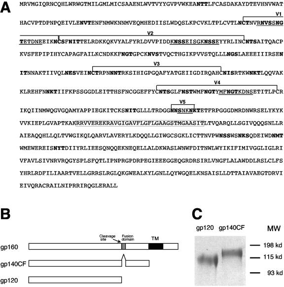
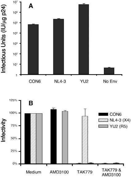
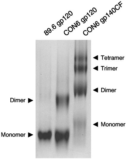
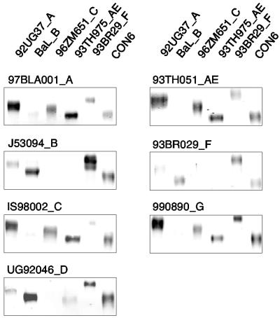
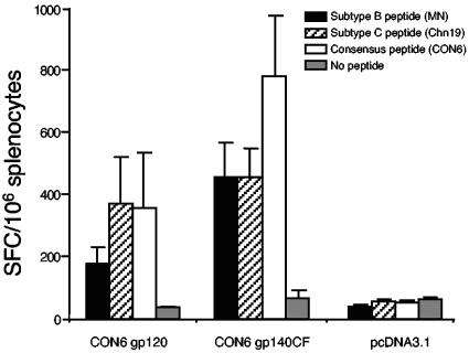
Genetic variation of human immunodeficiency virus (HIV-1) represents a major obstacle for AIDS vaccine development. To decrease the genetic distances between candidate immunogens and field virus strains, we have designed and synthesized an artificial group M consensus env gene (CON6 gene) to be equidistant from contemporary HIV-1 subtypes and recombinants. This novel envelope gene expresses a glycoprotein that binds soluble CD4, utilizes CCR5 but not CXCR4 as a coreceptor, and mediates HIV-1 entry. Key linear, conformational, and glycan-dependent monoclonal antibody epitopes are preserved in CON6, and the glycoprotein is recognized equally well by sera from individuals infected with different HIV-1 subtypes. When used as a DNA vaccine followed by a recombinant vaccinia virus boost in BALB/c mice, CON6 env gp120 and gp140CF elicited gamma interferon-producing T-cell responses that recognized epitopes within overlapping peptide pools from three HIV-1 Env proteins, CON6, MN (subtype B), and Chn19 (subtype C). Sera from guinea pigs immunized with recombinant CON6 Env gp120 and gp140CF glycoproteins weakly neutralized selected HIV-1 primary isolates. Thus, the computer-generated "consensus" env genes are capable of expressing envelope glycoproteins that retain the structural, functional, and immunogenic properties of wild-type HIV-1 envelopes.


Figures

FIG. 1.
FIG. 1.
Generation and expression of the group M consensus env gene (CON6). (A) The complete amino acid sequence of CON6 gp160 is shown. The five regions from the wild-type CRF08_BC (98CN006) env gene are indicated by underlined letters. Variable regions are indicated by brackets above the sequences. Potential N-liked glycosylation sites are highlighted with boldface letters. The deletion in gp140CF construct is shown in a box. (B) Constructs of CON6 gp120 and gp140CF. (C) Expression of CON6 gp120 and gp140CF. CON6 gp120 and gp140CF were purified from the cell culture supernatants of rVV-infected 293T cells with agarose Galanthus nivalis lectin columns. Both gp120 and gp140CF were separated on a 6% SDS-polyacrylamide gel under reducing conditions and stained with Coomassie blue.
FIG. 2.
FIG. 2.
Infectivity and coreceptor usage of CON6 envelope. (A) CON6 and control env plasmids were cotransfected with an HIV-1/SG3Δenv backbone into human 293T cells to generate Env pseudovirions. Culture supernatants were analyzed for p24 content and then used to infect JC53-BL cells (using serial fivefold dilutions). The infectivity was determined by counting the number of blue cells (infectious units) per microgram of p24 of pseudovirions after staining the infected cells for β-galactosidase expression. (B) Coreceptor usage of the CON6 env gene was determined on JC53-BL cells treated with AMD3100 and/or TAK799 for 1 h (37°C) and then infected with equal amounts of p24 (5 ng) of each Env pseudovirion. Infectivity in the control group (no blocking agent) was set as 100%. The blockage efficiency was expressed as the percentage of the infectivity of the virus in the absence of the blocking agent. Data shown are means ± standard deviations from three independent experiments.
FIG. 3.
FIG. 3.
BN-PAGE analysis of CON6 gp120 and gp140CF proteins. Affinity-purified 89.6 gp120, CON6 gp120, and CON6 gp140CF proteins were loaded onto a 3 to 8% Tris-acetate NuPAGE gel, and electrophoresis was carried out for 1.5 h at 150 V with 50 mM MOPS-50 mM Tris-HCl (pH 7.7)-0.03% Coomassie blue as the cathode running buffer and 50 mM MOPS-50 mM Tris-HCl (pH 7.7) as the anode buffer.
FIG. 4.
FIG. 4.
Binding of CON6 gp120 and gp140CF to sCD4 and anti-Env MAbs. (A and B) Each of the indicated MAbs and sCD4 were covalently immobilized to a CM5 sensor chip (BIAcore), and CON6 gp120 (A) or gp140CF (B) was injected over each surface (100 and 300 μg/ml, respectively). (C and D) To determine induction of 17b MAb binding to CON6 gp120 and gp140CF, CON6 gp120 (C) or gp140CF (D) proteins were captured (400 to 580 response units) on individual flow cells immobilized with sCD4 or MAb A32 or T8. Following stabilization of each of the surfaces, MAb 17b was injected and allowed to flow over each of the immobilized flow cells. (E) To determine binding of CON6 gp120 and gp140CF to human MAbs in ELISA, titers of stock solutions of 20 μg of MAbs 447-52D, F39F, A32, IgG1b12, and 2F5 were determined with CON6 gp120 and gp140CF glycoproteins. MAbs 447-52D (V3), F39F (V3) A32 (gp120), and IgG1b12 (CD4 binding site) each bound to both CON6 gp120 and gp140CF well, while 2F5 (anti-gp41 ELDKWAS) bound only CON6 gp140CF. The concentrations at the end point titer (end titer where the experimental versus the control value was ≥3.0) with gp120 for MAb 447-52D and F39F binding were <0.003 and 0.006 μg/ml, respectively; that for MAb A32 was <0.125 μg/ml; that for IgG1b12 was <0.002 μg/ml; and that for 2F5 with gp140CF was 0.016 μg/ml.
FIG. 5.
FIG. 5.
Western blot analysis of Env proteins of multiple subtypes against antisera of multiple subtypes. Equal amounts of Env proteins (100 ng) were separated on SDS-10% polyacrylamide gels (gp120: Bal, 96ZM651, 93TH975, and CON6; gp140: 92UG37, and 93BR029). Following electrophoresis, proteins were transferred to nitrocellulose membranes and reacted with sera from HIV-1-infected patients (1:1,000). Protein-bound antibody was probed with fluorescence-labeled secondary antibodies, and the images were scanned and recorded on an Odyssey infrared imager (Li-Cor, Lincoln, Nebr.). Subtypes are indicated by single letters after Env protein and serum designations. Four to six sera were tested for each subtype, and reaction patterns were similar among all sera from the same subtype. One representative result for each subtype serum is shown.
FIG. 6.
FIG. 6.
T-cell immune responses induced by CON6 Env immunogens in mice. Splenocytes were isolated from individual immunized mice (five mice per group). After splenocytes were stimulated in vitro with overlapping Env peptide pools of CON6, Chn19_C, MN_B, and medium, IFN-γ-producing cells were determined by the enzyme-linked immune spot assay. Total responses for each immunogen and peptide pool are expressed as SFCs per million splenocytes. The values are the means ± standard errors of the means of IFN-γ SFCs (n = 5 mice per group).

References

    1. Andre, S., B. Seed, J. Eberle, W. Schraut, A. Bultmann, and J. Haas. 1998. Increased immune response elicited by DNA vaccination with a synthetic gp120 sequence with optimized codon usage. J. Virol. 72:1497-1503. - PMC - PubMed
    1. Brandt, S. M., R. Mariani, A. U. Holland, T. J. Hope, and N. R. Landau. 2002. Association of chemokine-mediated block to HIV entry with coreceptor internalization. J. Biol. Chem. 277:17291-17299. - PubMed
    1. Bures, R., A. Gaitan, T. Zhu, C. Graziosi, K. M. McGrath, J. Tartaglia, P. Caudrelier, R. El Habib, M. Klein, A. Lazzarin, D. M. Stablein, M. Deers, L. Corey, M. L. Greenberg, D. H. Schwartz, and D. C. Montefiori. 2000. Immunization with recombinant canarypox vectors expressing membrane-anchored glycoprotein 120 followed by glycoprotein 160 boosting fails to generate antibodies that neutralize R5 primary isolates of human immunodeficiency virus type 1. AIDS Res. Hum. Retroviruses 16:2019-2035. - PubMed
    1. Bures, R., L. Morris, C. Williamson, G. Ramjee, M. Deers, S. A. Fiscus, S. Abdool-Karim, and D. C. Montefiori. 2002. Regional clustering of shared neutralization determinants on primary isolates of clade C human immunodeficiency virus type 1 from South Africa. J. Virol. 76:2233-2244. - PMC - PubMed
    1. Carr, J. K., B. T. Foley, T. Leitner, M. O. Salminen, B. T. Korber, and F. McCutchan. 1998. Reference sequences representing the principal genetic diversity of HIV-1 in the pandemic, p. III-10-III-19. In B. Korber, C. Kuiken, B. Foley, B. H. Hahn, F. McCutchan, J. W. Mellors, and J. Sodrosky (ed.), Human retroviruses and AIDS 1998: a compilation and analysis of nucleic acid and amino acid sequence. Theoretical Biology and Biophysics Group, Los Alamos National Laboratory, Los Alamos, N.Mex.

Publication types

MeSH terms

Substances