. 2005 Jan;79(2):1223-31.
doi: 10.1128/JVI.79.2.1223-1231.2005.

Variable surface epitopes in the crystal structure of dengue virus type 3 envelope glycoprotein

Affiliations

Variable surface epitopes in the crystal structure of dengue virus type 3 envelope glycoprotein

Yorgo Modis et al. J Virol. 2005 Jan.

Abstract

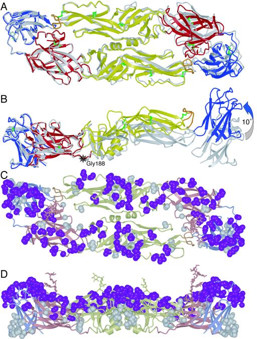
Dengue virus is an emerging global health threat. The major envelope glycoprotein, E, mediates viral attachment and entry by membrane fusion. Antibodies that bind but fail to neutralize noncognate serotypes enhance infection. We have determined the crystal structure of a soluble fragment of the envelope glycoprotein E from dengue virus type 3. The structure closely resembles those of E proteins from dengue type 2 and tick-borne encephalitis viruses. Serotype-specific neutralization escape mutants in dengue virus E proteins are all located on a surface of domain III, which has been implicated in receptor binding. While antibodies against epitopes in domain I are nonneutralizing in dengue virus, there are neutralizing antibodies that recognize serotype-conserved epitopes in domain II. The mechanism of neutralization for these antibodies is probably inhibition of membrane fusion. Our structure shows that neighboring glycans on the viral surface are spaced widely enough (at least 32 A) that they can interact with multiple carbohydrate recognition domains on oligomeric lectins such as DC-SIGN, ensuring maximum affinity for these putative receptors.


Figures

FIG. 1.
FIG. 1.
Structure of the sE dimer of dengue virus E sE in the mature virus particle. (A) The three domains of dengue virus sE. Domain I is red, domain II is yellow, and domain III is blue. A 53-residue stem segment links the stably folded sE fragment with the C-terminal transmembrane anchor. (B) The DEN-3 sE dimer viewed along its twofold symmetry axis. (C) The sE dimer viewed perpendicular to its twofold axis. The two glycans on residues 67 and 153 of the two subunits (A and B) of the dimer are labeled.
FIG. 2.
FIG. 2.
Contacts involving the glycans of sE between molecules related by the crystal symmetry. A closeup of the sE dimer (in red, yellow, and blue for domains I, II, and III, respectively) viewed from the side (inset) shows that both glycans form crystal contacts with an adjacent molecule in the crystal (shown in lighter colors). Mannose residues in the glycan on Asn-153 of chain A form hydrogen bonds with the main chain and side-chain oxygen atoms of Ser-16 and the side-chain amine of Lys-36. The N-acetylglucosamine linked to Asn-67 of chain B forms a hydrogen bond with a mannose in the glycan on Asn-153 of chain B of a neighboring molecule in the crystal.
FIG. 3.
FIG. 3.
Structure of the DEN-3 sE dimer superimposed on the DEN-2 sE dimer, using domain I from one monomer as the reference, viewed along the twofold symmetry axes (A) and perpendicular to the twofold axes (B). The greatest difference between the two structures is a ∼10° rotation of domain II relative to domain I about a point near Gly-188 (indicated by a grey star). (C) The DEN-3 sE dimer with residues that are not conserved in DEN-2 is shown in space-filling representation. Residues that are exposed on the viral surface are in magenta; residues that are not exposed are in grey. (D) The same structure as in panel C but viewed perpendicular to the twofold axis.
FIG. 4.
FIG. 4.
Antibody neutralization-escape mutants in dengue virus. (A) Three serotype-specific epitopes have been reported: residue 291 (2), residues 301 to 307 (29), and residues 381 to 383 (17, 18). Side chains in all three epitopes are shown on the DEN-3 E structure in magenta in space-filling representation. Changes at residue 388 (in pink), also unconserved and exposed, correlate with changes in virulence (28). Phe-277 (in green) is conserved in the dengue virus serotypes, but its mutation to serine during passaging in cell culture leads to escape from neutralization by IgM M10 (2, 27). (B) A side view of panel A is shown. (C) Stereoscopic view of a close-up of DEN-3 domain III, showing the four proposed serotype-specific epitopes. The orientation is halfway between the orientations used in the models shown in panels A and B, and the close-up is of the copy of domain III shown on the right in panels A and B. (D) The same view as in panel C is shown, but of DEN-2 (and with the homologous residues highlighted).

References

    1. Altmann, F., E. Staudacher, I. B. Wilson, and L. Marz. 1999. Insect cells as hosts for the expression of recombinant glycoproteins. Glycoconj. J. 16:109-123. - PubMed
    1. Beasley, D. W., and J. G. Aaskov. 2001. Epitopes on the dengue 1 virus envelope protein recognized by neutralizing IgM monoclonal antibodies. Virology 279:447-458. - PubMed
    1. Brandt, W. E., J. M. McCown, M. K. Gentry, and P. K. Russell. 1982. Infection enhancement of dengue type 2 virus in the U-937 human monocyte cell line by antibodies to flavivirus cross-reactive determinants. Infect. Immun. 36:1036-1041. - PMC - PubMed
    1. Brünger, A. T., P. D. Adams, G. M. Clore, W. L. DeLano, P. Gros, R. W. Grosse-Kunstleve, J. S. Jiang, J. Kuszewski, M. Nilges, N. S. Pannu, R. J. Read, L. M. Rice, T. Simonson, and G. L. Warren. 1998. Crystallography & NMR System: a new software suite for macromolecular structure determination. Acta Crystallogr. D Biol. Crystallogr. 54:905-921. - PubMed
    1. Burke, D. S., and T. P. Monath. 2001. Flaviviruses, p. 1043-1125. In D. M. Knipe, P. M. Howley, D. E. Griffin, S. R. A. Lamb, M. A. Martin, B. Roizman, and S. E. Straus (ed.), Fields virology, 4th ed. Lippincott Williams & Wilkins, Philadelphia, Pa.

Publication types

LinkOut - more resources