. 2005 Jan;79(2):1296-307.
doi: 10.1128/JVI.79.2.1296-1307.2005.

Terminal differentiation into plasma cells initiates the replicative cycle of Epstein-Barr virus in vivo

Affiliations

Terminal differentiation into plasma cells initiates the replicative cycle of Epstein-Barr virus in vivo

Lauri L Laichalk et al. J Virol. 2005 Jan.

Abstract

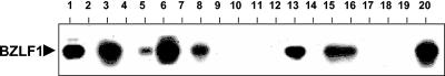
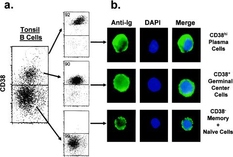
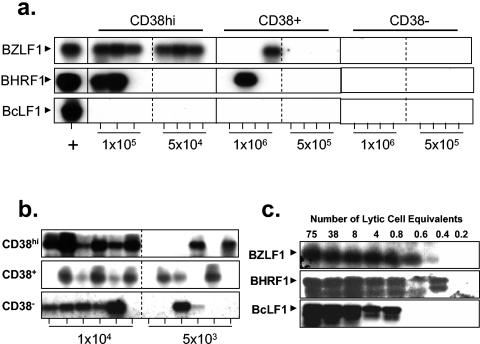
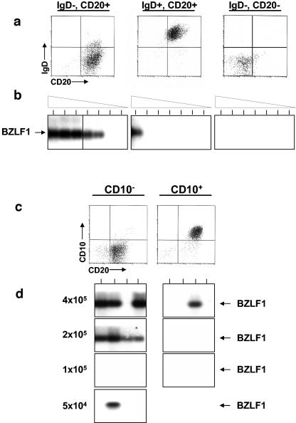
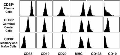
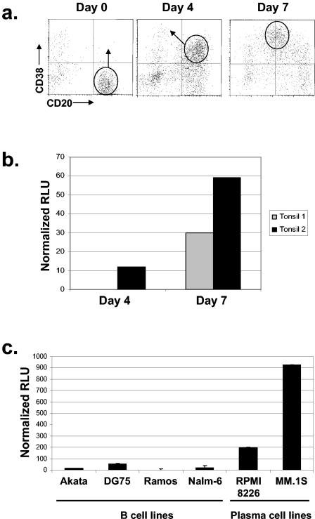
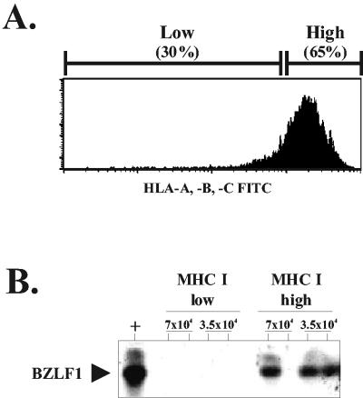
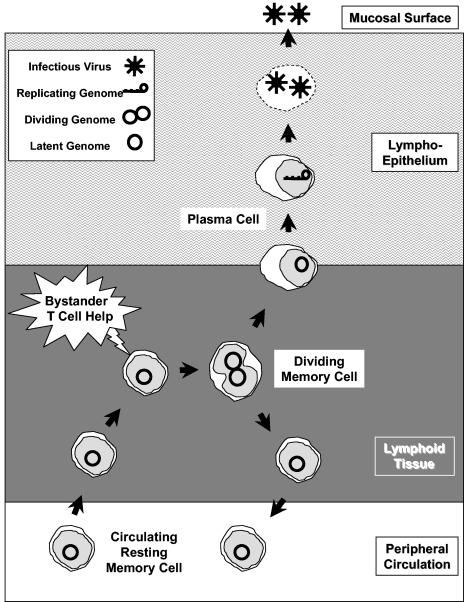
In this paper we demonstrate that the cells which initiate replication of Epstein-Barr virus (EBV) in the tonsils of healthy carriers are plasma cells (CD38hi, CD10-, CD19+, CD20lo, surface immunoglobulin negative, and cytoplasmic immunoglobulin positive). We further conclude that differentiation into plasma cells, and not the signals that induce differentiation, initiates viral replication. This was confirmed by in vitro studies showing that the promoter for BZLF1, the gene that begins viral replication, becomes active only after memory cells differentiate into plasma cells and is also active in plasma cell lines. This differs from the reactivation of BZLF1 in vitro, which occurs acutely and is associated with apoptosis and not with differentiation. We suggest that differentiation and acute stress represent two distinct pathways of EBV reactivation in vivo. The fraction of cells replicating the virus decreases as the cells progress through the lytic cycle such that only a tiny fraction actually release infectious virus. This may reflect abortive replication or elimination of cells by the cellular immune response. Consistent with the later conclusion, the cells did not down regulate major histocompatibility complex class I molecules, suggesting that this is not an immune evasion tactic used by EBV and that the cells remain vulnerable to cytotoxic-T-lymphocyte attack.


Figures

FIG. 1.
FIG. 1.
Screen of tonsils for replication of EBV. RNA was extracted from the unfractionated tonsil lymphocytes of healthy EBV carriers. RT-PCR was then performed for expression of BZLF1, the first gene expressed in the lytic cycle, which initiates the gene expression cascade leading to production of infectious virus. PCR products were identified by Southern blotting with a BZLF1-specific probe.
FIG. 2.
FIG. 2.
Flow cytometric fractionation of tonsil B lymphocytes based on expression of CD38. (a) Tonsil lymphocytes were depleted of T cells by using MACS beads and the CD3 monoclonal antibody. The remaining B cells were stained with CD38. Three discrete populations were observed and separated with a Mo-Flo (Dako): CD38hi (plasma cells), CD38+ (germinal center cells), and CD38 (naive and memory cells). (b) The CD38hi B cells are plasma cells. The sorted cells from panel a were stained for immunoglobulin expression (green), and the nucleus was identified by DAPI staining (blue). Only the CD38hi subset consisted of plasma cells, based on high-level expression of cytoplasmic immunoglobulin, lack of membrane immunoglobulin, a large cytoplasm-to-nucleus ratio, and a displaced nucleus.
FIG. 3.
FIG. 3.
EBV replicates in the CD38hi subsets of tonsil B cells. CD38hi, CD38+, and CD38 B cells were separated by FACS from a single tonsil, subjected to limiting-dilution analyses, and analyzed for the presence of viral RNA and DNA. (a) The cell populations were tested by RT-PCR for expression of an immediate-early (BZLF1), an early (BHRF1), and a late (BcLF1) replicative gene. Due to the relatively low yields of pure CD38hi plasma cells, 10-fold higher numbers of cells could be tested for the CD38+ and CD38 subsets. (b) The cell populations were also tested for the presence of the virus by DNA PCR. PCR products were identified by Southern blotting with a gene-specific probe. Tonsil 1 from Table 1 was used in this experiment. (c) Sensitivity of the RT-PCR assays for EBV replicative genes. Akata cells were induced into the lytic cycle by cross-linking of the surface immunoglobulin. cDNA was then prepared, and serial dilutions were tested by RT-PCR analysis for detection of the immediate-early (BZLF1), early (BHRF1), and late (BcLF1) replicative genes. The fraction of cells replicating the virus in the cultures was estimated by immunofluorescence staining for the BZLF1 protein.
FIG. 4.
FIG. 4.
EBV replicates in the IgD CD10 CD20+ subset of tonsil B cells. (a) T-cell-depleted tonsil lymphocytes (CD3) were fractionated by MACS into IgD+ and IgD fractions. The IgD subset was further fractionated by MACS into CD20+ and CD20 fractions. The purified cells were analyzed by flow cytometry for purity (top panels). (The T-cell depletion was less efficient for this experiment than for the one shown in Fig. 2.) The CD20 fraction probably consists of contaminating T cells and macrophages but could also include plasma cells that had lost their lineage markers. (b) Cells (106) of each fraction were subjected to serial twofold dilutions, and the dilutions were tested by RT-PCR for expression of the immediate-early gene BZLF1. PCR products were identified by Southern blotting with a gene-specific probe. Tonsil 1 from Table 1 was used in this experiment. (c) CD10+ CD20+ and CD10 CD20+ tonsil lymphocytes were separated by FACS sorting. (d) The samples were serially diluted, and then multiple replicates were made for each dilution. Each sample was analyzed by RT-PCR (upper panels) for expression of the immediate-early BZLF1 gene. PCR products were identified by Southern blotting with a gene-specific probe. Tonsil 2 from Table 1 was used in this experiment.
FIG. 5.
FIG. 5.
Cell surface phenotyping of the three populations separated on the basis of CD38 expression. CD38hi, CD38+, and CD38 B cells were analyzed by flow cytometry for expression of CD38, the pan-B-cell markers CD19 and CD20, CD138 (Syndecan), MHC class I, and the germinal center-specific marker CD10. Isotype-matched antibodies were used for the negative controls. The plasma cells are CD38hi B cells that express reduced levels of CD20 and are CD138 (Syndecan) negative.
FIG. 6.
FIG. 6.
The BZLF1 promoter Zp functions in plasma cells but not activated memory cells. (a) Resting tonsil memory cells (CD38 CD20+) were isolated by negative selection and induced to differentiate by culturing with IL-2 and IL-10 together with NIH 3T3 fibroblasts expressing CD40 ligand as described previously (3). After 4 days of culture a subset of cells had adopted the phenotype of activated memory cells (CD38+ CD20+) (10), and after 7 days they had adopted that of plasma cells (CD38hi CD20lo). (b) Relative transcriptional activity of a BZLF1 promoter-luciferase reporter construct transfected into resting memory B cells after 4 or 7 days of culture as described for panel a. The results of two separate experiments are shown. (c) As for panel b, but with a collection of human B-cell lines, including two plasma cell lines. Error bars indicate standard deviations. RLU, Relative light units.
FIG. 7.
FIG. 7.
MHC class I expression is not down regulated on plasma cells replicating EBV. Plasma cells were purified as described in the legend to Fig. 2. The cells were then stained for expression of MHC class I molecules and separated into low and high expressers as shown. The two populations were then subjected to RT-PCR to detect expression of the viral BZLF1 gene as described in the legend to Fig. 3.
FIG. 8.
FIG. 8.
A model of how EBV may persist in the circulating memory B-cell compartment while being continuously shed into saliva, based on the work of Bernasconi et al (7). They have suggested that memory B cells exposed to bystander T-cell help become activated and divide, generating new memory cells and plasma cells. This process maintains the level of memory cells and antibody-secreting plasma cells in an antigen-independent way. If the memory B cell contained latent EBV, the process would produce another latently infected memory cell, maintaining the levels of latently infected cells in the peripheral circulation, while also producing a plasma cell, thus also ensuring the continuous release of infectious virus. Since this process occurs in the tonsils, the plasma cell would migrate into the lymphoepithelium and produce the virus. Whether this virus is released directly into saliva or is first amplified through lytic infection of epithelial cells is a controversial and, as yet, unresolved issue.

References

    1. Adler, B., E. Schaadt, B. Kempkes, U. Zimber-Strobl, B. Baier, and G. W. Bornkamm. 2002. Control of Epstein-Barr virus reactivation by activated CD40 and viral latent membrane protein 1. Proc. Natl. Acad. Sci. USA 99:437-442. - PMC - PubMed
    1. Anagnostopoulos, I., M. Hummel, C. Kreschel, and H. Stein. 1995. Morphology, immunophenotype, and distribution of latently and/or productively Epstein-Barr virus-infected cells in acute infectious mononucleosis: implications for the interindividual infection route of Epstein-Barr virus. Blood 85:744-750. - PubMed
    1. Arpin, C., J. Banchereau, and Y. J. Liu. 1997. Memory B cells are biased towards terminal differentiation: a strategy that may prevent repertoire freezing. J. Exp. Med. 186:931-940. - PMC - PubMed
    1. Babcock, G. J., L. L. Decker, M. Volk, and D. A. Thorley-Lawson. 1998. EBV persistence in memory B cells in vivo. Immunity 9:395-404. - PubMed
    1. Babcock, G. J., D. Hochberg, and A. D. Thorley-Lawson. 2000. The expression pattern of Epstein-Barr virus latent genes in vivo is dependent upon the differentiation stage of the infected B cell. Immunity 13:497-506. - PubMed

Publication types

LinkOut - more resources