Comparative Study
. 2005 Mar;88(3):1932-47.
doi: 10.1529/biophysj.104.044412. Epub 2004 Dec 21.

The photophysics of green fluorescent protein: influence of the key amino acids at positions 65, 203, and 222

Affiliations
Comparative Study

The photophysics of green fluorescent protein: influence of the key amino acids at positions 65, 203, and 222

Gregor Jung et al. Biophys J. 2005 Mar.

Abstract

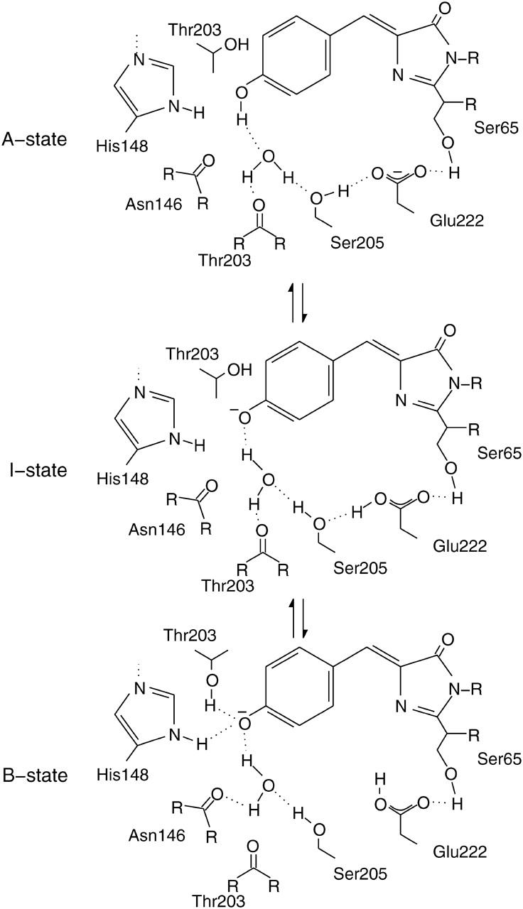
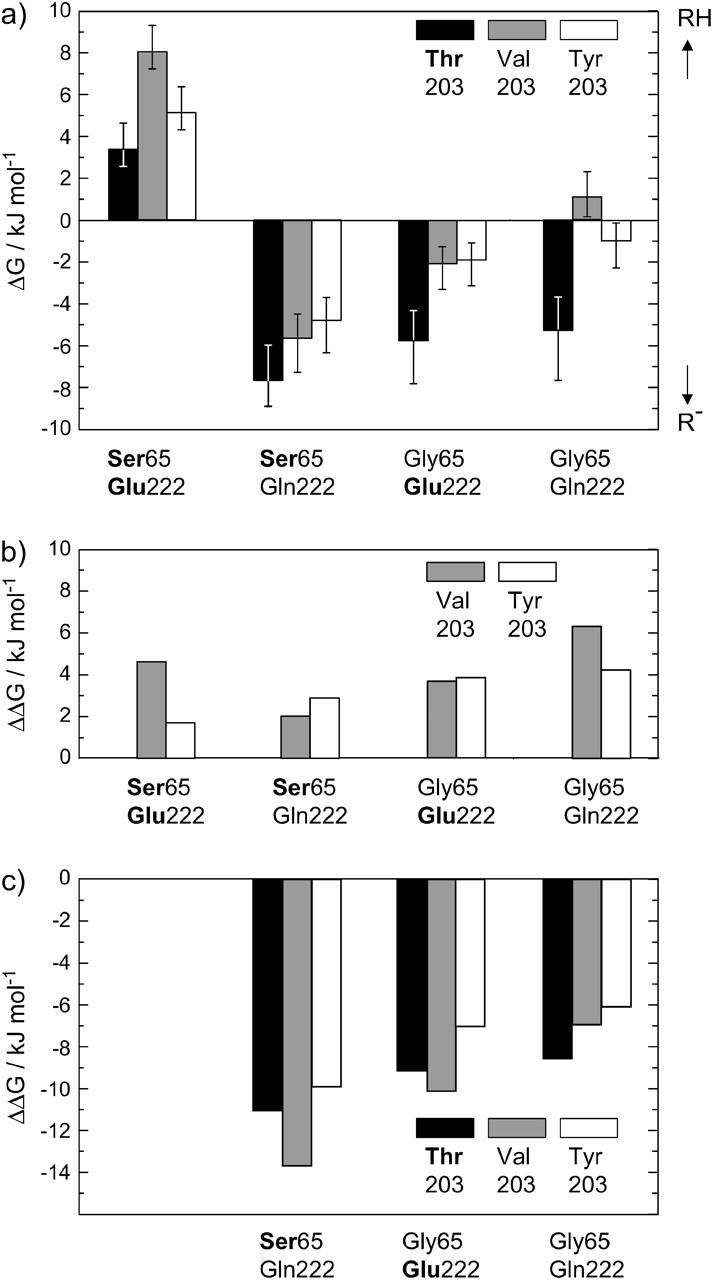
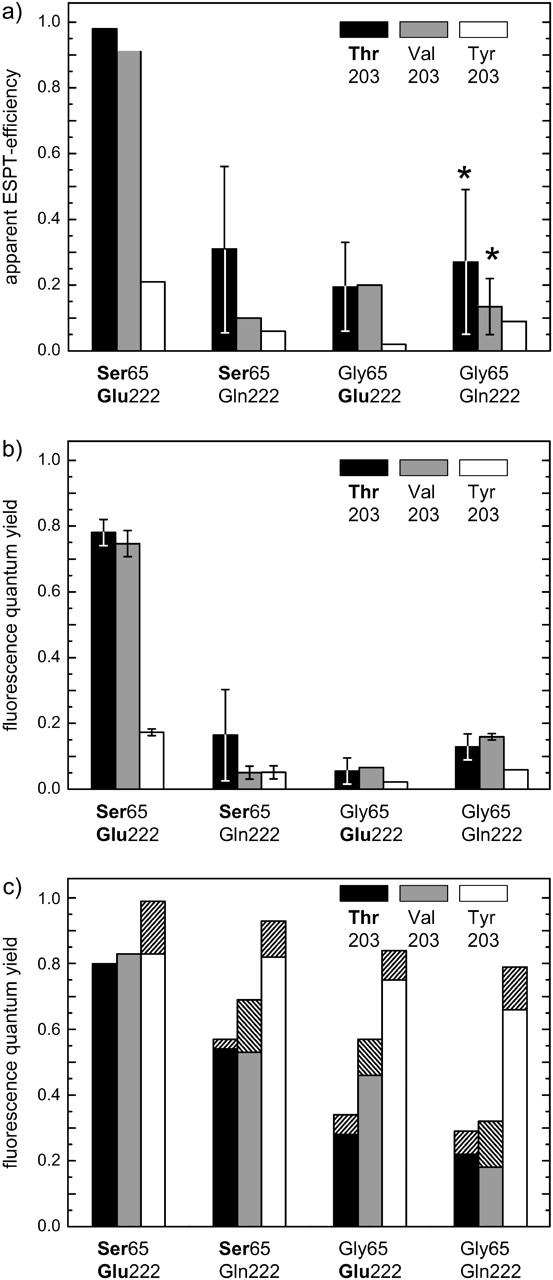
The three amino acids S65, T203, and E222 crucially determine the photophysical behavior of wild-type green fluorescent protein. We investigate the impact of four point mutations at these positions and their respective combinations on green fluorescent protein's photophysics using absorption spectroscopy, as well as steady-state and time-resolved fluorescence spectroscopy. Our results highlight the influence of the protein's hydrogen-bonding network on the equilibrium between the different chromophore states and on the efficiency of the excited-state proton transfer. The mutagenic approach allows us to separate different mechanisms responsible for fluorescence quenching, some of which were previously discussed theoretically. Our results will be useful for the development of new strategies for the generation of autofluorescent proteins with specific photophysical properties. One example presented here is a variant exhibiting uncommon blue fluorescence.


Figures

FIGURE 1
FIGURE 1
Model of the GFP chromphore in its three different states, A, B, and I (Brejc et al., 1997). Mutations in the surrounding hydrogen-bonding network influence the equilibrium between these states and are therefore responsible for spectral shifts observed in the absorbance and fluorescence spectra.
FIGURE 2
FIGURE 2
Absorption (black line) and fluorescence spectra for the GFP variants under investigation. All data are recorded in aqueous buffer solution at room temperature with protein concentrations of 5–6 μM. Fluorescence emission spectra after excitation of the RH form (green line) were recorded with an excitation wavelength of λexc = 380 nm. For the recording of fluorescence emission spectra after excitation of R form (blue line), λexc values of 5–15 nm below the maximum of the R form were chosen. Fluorescence excitation spectra (red line) were recorded with a detection wavelength λdet = 530 nm for the variants carrying the mutations T203 and T203V and λdet = 540 nm for the variants with the mutation T203Y. To illustrate different fluorescence quantum yields for RH and R forms, the excitation spectra were normalized to the maxima of the R forms in the absorption spectra. Bold print denotes the amino acids of wt-GFP.
FIGURE 3
FIGURE 3
(a) ΔG values for the different chromophore states in the investigated GFP mutants, calculated from the equilibrium constant K. The error bars result from an assumed error of ±40% in the determination of K. An additional error of 50% is included for the mutants with weak RH-absorbance. Bold print denotes the amino acids of wt-GFP. (b) Energy values ΔΔG relative to the T203 variants. The removal of the hydrogen bond between Y66 and the amino acid at position 203 stabilizes the neutral chromophore. (c) Energy values ΔΔG relative to the S65/E222 variants. The perturbation of the hydrogen-bonding network between Y66 and E222 favors the deprotonated chromophore form.
FIGURE 4
FIGURE 4
Excited-state proton transfer efficiences and quantum yields derived from the spectral data. (a) ESPT efficiencies in the GFP variants. The asterisk denotes the variants with strong blue RH fluorescence, which prevents an accurate determination of the ESPT quantum yield. Low quantum yield values might be caused by an unavoidable blue-edge excitation of the deprotonated form, by a subconformation with fast ESPT in the S65G/E222-variants like in EGFP (Cotlet et al., 2001), or by an alternative ESPT-mechanism (McAnaney et al., 2002; Winkler et al. 2002). The large error bars in a result from the weak absorbance of the neutral form or from the strong overlap of the RH and R fluorescence emissions. (b) Fluorescence quantum yields ΦFl for the neutral form referenced to the deprotonated form in wt-GFP with ΦFl = 0.8. The large error bars in b result from weak absorbance of the neutral form. (c) Fluorescence quantum yields for the deprotonated form referenced to wt-GFP with ΦFl = 0.8. The values obtained by the Strickler-Berg relation are indicated by hatched bars (Strickler and Berg, 1962).
FIGURE 5
FIGURE 5
Time-correlated single-photon counting measurements with excitation wavelengths between λexc = 385–390 nm performed to reveal the origin of the blue fluorescence. (a) A rising, isotope-sensitive component in the green fluorescence of T203V detected at λdet = 515 nm indicates ESPT. The rising components are fit by a single exponential. (b) Fluorescence lifetime measurements of S65G/T203V/E222Q with λdet = 460 nm in H2O and D2O. Similar curves are obtained for S65G/E222Q (data not shown). (c) Fluorescence lifetime measurements of S65G/T203V/E222Q with λdet = 515 nm in H2O and D2O. Similar curves are obtained for S65G/E222Q (data not shown). (d) Lifetime measurements in S65G/E222Q at 2 K with λexc = 420 nm. Neither a rising component at 503 nm nor a fast-decaying component at 445 nm is seen.
FIGURE 6
FIGURE 6
Spectral properties of GFP variants before and after photoconversion. (a) Absorption spectra of the T203V mutant. The original absorption spectrum (black line) is only slightly affected by illumination with near-UV light (350–450 nm) from the arc lamp for 1 h (shaded line). Photoconversion is induced with an additional 2 min of illumination with the integral light of the arc lamp (dotted line). (b) Absorption spectra of S65G/T203Y before (solid line) and after (dotted line) photoconversion. Reversed photoconversion of the deprotonated chromophore form is observed with UV illumination (2 min with the integral arc lamp emission). S65G and S65G/T203V behave similarily (data not shown). (c) Comparison between the fluorescence lifetime of the deprotonated chromophore form in S65G/T203V before (black line) and after (shaded line) photoconversion. Excitation and detection wavelengths were chosen as λexc = 465–470 nm and λdet = 515 nm, respectively. The faster fluorescence decay indicates a higher contribution of internal conversion in the photoproducts (cf. Table 4).
FIGURE 7
FIGURE 7
Chromophore forms and internal conversion pathways. (a) Benzoidal and quinoidal mesomers of the GFP chromophore in its deprotonated form. The benzoidal character is increased if the negative charge on the oxygen of Y66 is stabilized by hydrogen bonds. The benzoidal structure prevails in the neutral chromophore form. (b) The one-bond rotation around the angle φ. With increasing single-bond character of φ, this movement becomes likelier. In contrast, increasing quinoidal character as in the I-state reduces its contribution to internal conversion. (c) The hula-twist motion, which is less sensitive to the bond-order of the exocyclic bonds. However, this concerted two-bond rotation requires space near the heterocycle and is therefore probed by S65G mutations and photoconversion.

References

    1. Bae, J. H., M. Rubini, G. Jung, G. Wiegand, M. H. J. Seifert, M. K. Azim, J. S. Kim, A. Zumbusch, T. Holak, L. Moroder, R. Huber, and N. Budisa. 2003. Expansion of the genetic code enables design of a novel “gold” class of green fluorescent proteins. J. Mol. Biol. 328:1071–1081. - PubMed
    1. Baffour-Awuah, N., and M. Zimmer. 2004. Hula-twisting in green fluorescent protein. Chem. Phys. 303:7–11.
    1. Barondeau, D. P., C. D. Putnam, C. J. Kassmann, J. A. Tainer, and E. D. Getzoff. 2003. Mechanism and energetics of green fluorescent protein chromophore synthesis revealed by trapped intermediate structures. Proc. Natl. Acad. Sci. USA. 100:12111–12116. - PMC - PubMed
    1. Bell, A. F., D. Stoner-Ma, R. M. Wachter, and P. J. Tonge. 2003. Light-driven decarboxylation of wild-type green fluorescent protein. J. Am. Chem. Soc. 125:6919–6926. - PubMed
    1. Branchini, B. R., A. R. Nemser, and M. Zimmer. 1998. A computational analysis of the unique protein-induced tight turn that results in posttranslational chromophore formation in green fluorescent protein. J. Am. Chem. Soc. 120:1–6.

Publication types

MeSH terms

Substances

LinkOut - more resources