. 2005 Jan;89(1):30-5.
doi: 10.1136/bjo.2003.040543.

Does visual restitution training change absolute homonymous visual field defects? A fundus controlled study

Affiliations

Does visual restitution training change absolute homonymous visual field defects? A fundus controlled study

J Reinhard et al. Br J Ophthalmol. 2005 Jan.

Abstract

Aim: To examine whether visual restitution training (VRT) is able to change absolute homonymous field defect, assessed with fundus controlled microperimetry, in patients with hemianopia.

Methods: 17 patients with stable homonymous visual field defects before and after a 6 month VRT period were investigated with a specialised microperimetric method using a scanning laser ophthalmoscope (SLO). Fixation was controlled by SLO fundus monitoring. The size of the field defect was quantified by calculating the ratio of the number of absolute defects and the number of test points; the training effect E was defined as the difference between these two ratios before and after training. A shift of the entire vertical visual field border by 1 degrees would result in an E value of 0.14.

Results: The mean training effect of all right eyes was E = 0.025 (SD 0.052) and all left eyes E = 0.008 (SD 0.034). In one eye, a slight non-homonymous improvement along the horizontal meridian occurred.

Conclusions: In one patient, a slight improvement along the horizontal meridian was found in one eye. In none of the patients was an explicit homonymous change of the absolute field defect border observed after training.


Figures

Figure 5
Figure 5
Figure 1
Figure 1
Stimulus presentation in the SLO. The investigator sees stimuli and retina simultaneously. When fixation is central—that is, the foveola was located on the fixation cross, he presented a test triplet (120 ms presentation time). In (A) a triplet with 3° eccentricity and 2° inter-dot distance is shown. The patient’s task was to say how many and which of the dots he could see (for example, the upper, the lower, and/or the middle). Eccentricity and inter-dot distance were varied in random order; altogether a grid of 241 locations in central visual field was tested (B). The grid covered an area ranging 1° towards the healthy field and 10° in the blind field, vertically plus or minus 8°.
Figure 2
Figure 2
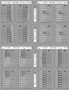
Original findings of SLO specialised microperimetry of the central visual field in four patients, before and after training. The white dots were perceived by the patients, the black dots were not. The broken black lines represent the horizontal and vertical meridians. Patient 13 has a complete homonymous hemianopia to the left side without macular sparing. After training, no substantial change in the absolute field defect was seen. In the left eye a slight torsion occurred. Patient 17 has a small homonymous paracentral scotoma that remained unchanged after the training. Patient 5 presents with a homonymous incomplete quadrantanopia to the upper right side. The absolute field defect decreased its size along the horizontal meridian, especially in the right eye. However, this improvement is not congruent and no substantial change occurred along the vertical meridian. Patient 8 has a complete hemianopia to the right with a vertical strip of perception along the vertical meridian. In the left eye, a slight improvement of the visual field occurred (EOS = 0.029), the visual field of the right eye remained unchanged (EOD = 0.0).
Figure 3
Figure 3
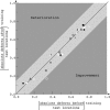
Results of the quantitative evaluation of the SLO microperimetry of all patients. White symbols represent right, black symbols left eyes. Data points on the identity line symbolise no change in the absolute defects (E = 0), data points above this line show a deterioration, those below an improvement. Data points within the light grey area around the identity line represent eyes with a change of the entire border of less than one degree (E<0.14). With exception of the results of the right eye of patient 5, all eyes are located within this area. Only the right eye of patient 5 is located outside this area (see also fig 2 and table 1).
Figure 4
Figure 4
Reading speeds (in words per minute) before and after training. Data points on the identity line represent no change in reading speed after training, data points within the light grey area represent a change of less than 20% of the reading speed after training. Three patients improved more than 20% with regard to reading speed.

Comment in

  • Vision restoration therapy.
    Sabel BA, Kenkel S, Kasten E. Sabel BA, et al. Br J Ophthalmol. 2005 May;89(5):522-4. doi: 10.1136/bjo.2005.068163. Br J Ophthalmol. 2005. PMID: 15834073 Free PMC article. No abstract available.
  • Vision restoration therapy: confounded by eye movements.
    Horton JC. Horton JC. Br J Ophthalmol. 2005 Jul;89(7):792-4. doi: 10.1136/bjo.2005.072967. Br J Ophthalmol. 2005. PMID: 15965150 Free PMC article. No abstract available.
  • Vision restoration therapy.
    Caplan LR, Firlik A, Newman NJ, Pless M, Romano JG, Schatz N. Caplan LR, et al. Br J Ophthalmol. 2005 Sep;89(9):1229. doi: 10.1136/bjo.2005.069773. Br J Ophthalmol. 2005. PMID: 16113396 Free PMC article. No abstract available.

References

    1. Kasten E , Wüst S, Behrens-Baumann W, et al. Computer-based training for the treatment of partial blindness. Nat Med 1998;4:1083–7. - PubMed
    1. Sabel BA, Kasten E. Restoration of vision by training of residual functions. Curr Opin Ophthalmol 2000;11:430–6. - PubMed
    1. Kasten E , Wüst S, Sabel BA. Residual vision in transition zones in patients with cerebral blindness. J Clin Exp Neuropsychol 1998;20:581–98. - PubMed
    1. Kommerell G , Lieb B, Münssinger U. Rehabilitation bei homonymer Hemianopsie. Z prakt Augenheilkd 1999;20:344–52.
    1. Trauzettel-Klosinski S , Reinhard J. The vertical field border in hemianopia and its significance for fixation and reading. Invest Ophthalmol Vis Sci 1998;39:2177–86. - PubMed