. 2005 Jan;89(1):74-80.
doi: 10.1136/bjo.2004.051540.

Subthreshold diode micropulse photocoagulation for the treatment of clinically significant diabetic macular oedema

Affiliations

Subthreshold diode micropulse photocoagulation for the treatment of clinically significant diabetic macular oedema

J K Luttrull et al. Br J Ophthalmol. 2005 Jan.

Abstract

Aim: To report the visual and clinical outcomes of a pilot study of subthreshold diode micropulse (SDM) laser photocoagulation for clinically significant diabetic macular oedema (CSMO).

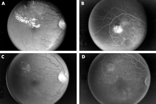
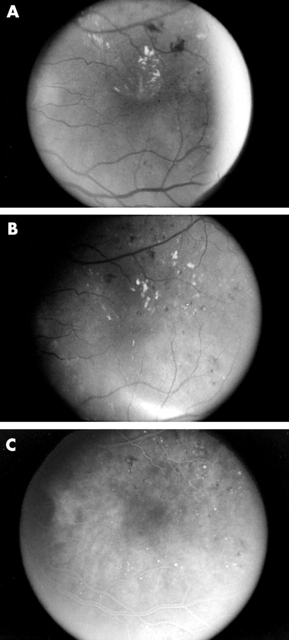
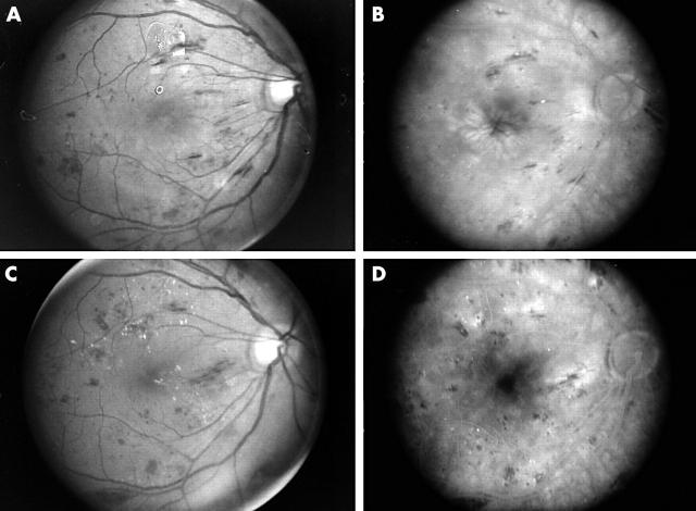
Methods: The results of infrared (810 nm) SDM laser photocoagulation for CSMO were retrospectively reviewed in 95 eyes of 69 consecutive patients with mild to moderate non-proliferative diabetic retinopathy. The same laser parameters were used for each patient. Only the number of laser applications varied between patients, depending on their macular findings. Primary outcome measures were Snellen visual acuity, fluorescein angiographic leakage, and CSMO status.

Results: Visual acuity was stable or improved in 85% of treated eyes, with a mean follow up of 12.2 months (range 3-29 months). CSMO decreased in 96% and resolved in 79% of treated eyes. No adverse laser events occurred. No laser lesions were detectable ophthalmoscopically or angiographically after treatment, consistent with calculations based on ANSI Z136.1 laser safety standards suggestive of only histologically detectable tissue effects at the laser exposure levels. No laser scarring was observed during the follow up period.

Conclusion: Subthreshold diode micropulse laser photocoagulation minimises chorioretinal damage in the management of CSMO and demonstrates a beneficial effect on visual acuity and CSMO resolution. Prospective studies are needed to fully evaluate this technique.


Figures

Figure 1
Figure 1
(A) Patient 206. Preoperative red-free fundus photograph demonstrating clinically significant diabetic macular oedema. (B) Preoperative intravenous fundus fluorescein angiogram. Note prominent focal retinal microvascular leakage. This patient was treated confluently throughout the area of leakage and retinal thickening with 269 applications of SDM photocoagulation. (C) Red-free fundus photograph 10 months following SDM photocoagulation. Note resolution of macular oedema and hard exudates. (D) Intravenous fundus fluorescein angiogram 10 months post-SDM macular photocoagulation. Note persistent but diminished focal microvascular leakage, and absence of angiographically visible pigment disturbance or chorioretinal scarring.
Figure 2
Figure 2
(A) Patient 141. Preoperoperative red-free fundus photograph demonstrating clinically significant diabetic macular oedema. (B) Red-free fundus photograph 5 months following 1663 applications of SDM photocoagulation applied confluently to the areas of biomicroscopically visible retinal thickening. The clinically significant macular oedema has resolved. (C) Postoperative intravenous fundus fluorescein angiogram. Note the absence of angiographically visible pigment disturabance or chorioretinal scarring following SDM macular photocoagulation.
Figure 3
Figure 3
(A) Patient 107. Preoperative red-free fundus photograph of patient with diffuse clinically significant diabetic macular oedema and foveal cysts. (Note film development artefacts superior to fovea and at temporal edge of photograph.) (B) Late phase preoperative intravenous fundus fluorescein angiogram of diffuse clinically significant diabetic macular oedema. Note cystoid leakage pattern in fovea. This patient was treated with 602 applications of SDM photocoagulation in a nearly confluent grid pattern throughout the macula extending to the edge of the fovea circumferentially. (C) Red-free fundus photograph 8 months following SDM photocoagulation. Note marked reduction in macular oedema without visible chorioretinal scarring or pigmentary disturbance. (D) Postoperative intravenous fundus fluorescein angiogram. Note marked reduction in diffuse and cystoid leakage. Note absence of angiographically visible pigmentary disturbance or chorioretinal scarring following SDM macular photocoagulation.

References

    1. Kahn HA, Hiller R. Blindness caused by diabetic retinopathy. Am J Ophthalmol 1974;78:58–67. - PubMed
    1. Kahn HA, Bradley RF. Prevalence of diabetic retinopathy. Age, sex, and duration of diabetes. Br J Ophthalmol 1975;59:345–9. - PMC - PubMed
    1. Aiello LM, Rand LI, Briones JC, et al. Diabetic retinopathy in Joslin Clinic patients with adult-onset diabetes. Ophthalmology 1981;88:619–23. - PubMed
    1. Klein R , Klein BE, Moss SE, et al. The Wisconsin Epidemiologic Study of diabetic retinopathy. XIV. Ten-year incidence and progression of diabetic retinopathy. Arch Ophthalmol 1994;112:1217–28. - PubMed
    1. McMeel JW, Trempe CL, Franks EB. Diabetic maculopathy. Trans Am Acad Ophthalmol Otolaryngol 1977;83 (Pt 1) :OP476–87. - PubMed

MeSH terms