. 2005 Jan 15;118(Pt 2):291-300.
doi: 10.1242/jcs.01623. Epub 2004 Dec 22.

A specific alpha5beta1-integrin conformation promotes directional integrin translocation and fibronectin matrix formation

Affiliations

A specific alpha5beta1-integrin conformation promotes directional integrin translocation and fibronectin matrix formation

Katherine Clark et al. J Cell Sci. .

Abstract

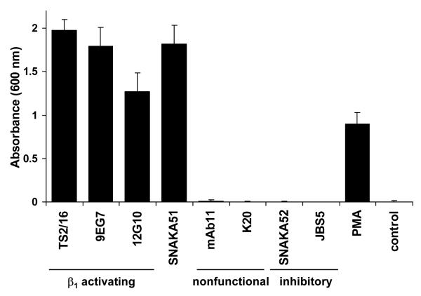
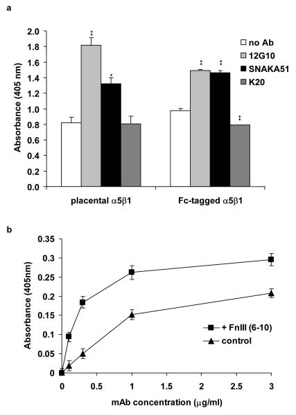
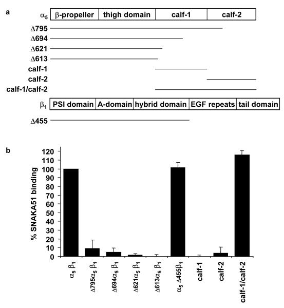
Integrin adhesion receptors are structurally dynamic proteins that adopt a number of functionally relevant conformations. We have produced a conformation-dependent anti-alpha5 monoclonal antibody (SNAKA51) that converts alpha5beta1 integrin into a ligand-competent form and promotes fibronectin binding. In adherent fibroblasts, SNAKA51 preferentially bound to integrins in fibrillar adhesions. Clustering of integrins expressing this activation epitope induced directional translocation of alpha5beta1, mimicking fibrillar adhesion formation. Priming of alpha5beta1 integrin by SNAKA51 increased the accumulation of detergent-resistant fibronectin in the extracellular matrix, thus identifying an integrin conformation that promotes matrix assembly. The SNAKA51 epitope was mapped to the calf-1/calf-2 domains. We propose that the action of the antibody causes the legs of the integrin to change conformation and thereby primes the integrin to bind ligand. These findings identify SNAKA51 as the first anti-integrin antibody to selectively recognize a subset of adhesion contacts, and they identify an integrin conformation associated with integrin translocation and fibronectin matrix formation.


Figures

Figure 1
Figure 1. K562 cell adhesion to fibronectin is promoted by SNAKA51 and other stimulatory anti-β1 antibodies
K562 cells were allowed to attach to a fibronectin-coated surface (2 μg/ml) in the presence of the indicated anti-integrin antibodies (10 μg/ml), PMA (100 nM), or no antibody (control). Unattached cells were removed, and remaining cells were fixed and stained with crystal violet. Cell attachment was quantified by absorbance measured at 600 nm.
Figure 2
Figure 2. SNAKA51 promotes ligand-binding to α5β1 integrin, and SNAKA51 binding to α5β1 integrin is increased in the presence of ligand
Using a solid-phase ligand-binding assay, the binding of biotinylated FnIII (6-10) (0.1 μg/ml) to α5β1 integrin was measured. a) Binding of FnIII (6-10) to directly coated placental α5β1 integrin and anti-Fc captured recombinant integrin in the presence and absence of SNAKA51, 12G10, or K20. Statistical analysis was performed using a 2-tailed t-test in comparison to the no antibody control. * p<0.005, **p<0.0005. b) Dose-dependent binding of biotinylated SNAKA51 to K20 tethered placental α5β1 integrin in the presence or absence of FnIII (6-10) (20 μg/ml).
Figure 3
Figure 3. SNAKA51 only colocalizes with fibronectin fibers and primed β1 integrin in fibrillar adhesions
Human fibroblasts were fixed and stained using indirect immunofluorescence for: a) α5 integrin [SNAKA51 (left panels) or SNAKA52 (center panels) or mAb11 (right panels), green], fibronectin (blue) and αV integrin as a focal adhesion marker (L230, red); or b) Top panel β1 integrin in a primed conformation (9EG7, red) and α5 integrin (SNAKA51, green), middle panel total β1 integrin (mAb11 red) and α5 integrin (SNAKA51, green), bottom panel β1 integrin in a primed conformation (9EG7, red) and total β1 integrin (mAb11 green). Bar 20 μm, insert bar 5μm.
Figure 4
Figure 4. The SNAKA51 epitope maps to the calf domains of the α5 subunit
a) Diagrammatic representation of the extracellular domains of the α5 and β1 integrin subunits indicating the position of truncations for the various recombinant constructs used in this study. b) The SNAKA51 binding site on the α5 integrin subunit was mapped using a sandwich ELISA of Fc-tagged truncated or individual domains of recombinant integrin. The Fc-tagged protein was specifically captured using anti-Fc antibodies, and then binding of SNAKA51 to the integrin was measured using enzyme-conjugated anti-mouse secondary antibody.
Figure 5
Figure 5. Induction of priming by SNAKA51 is dependent on the β1 leg and modulates β1 A-domain ligand-binding ability
Binding of biotinylated FnIII (6-10) to either wild-type or ADMIDAS mutant (D138A) recombinant α5β1 integrin, containing either a full-length or truncated (Δ455) β1 subunit, was measured in the presence or absence of anti-integrin antibodies.
Figure 6
Figure 6. Clustering of SNAKA51-bound α5 integrin induces integrin translocation out of focal adhesions and across the cell surface
Fibroblasts were plated on glass coverslips and incubated overnight in fibronectin-depleted medium with cycloheximide. Cells were labelled with antibody for 20 minutes, using SNAKA51 (a), SNAKA52 (b), or mAb11 (c). Unbound antibody was rinsed away, and the cells were either fixed (‘no chasing’), or the cell-bound antibody was clustered with goat anti-mouse or anti-rat IgG and incubated for a further 30 minutes (‘30’ chasing’) before fixation. For unclustered α5 integrin, cells were stained with anti-mouse or anti-rat IgG (green). For clustered α5 integrin, cells were stained with anti-goat IgG (green). In addition, all samples were stained with anti-αV antibody L230 (red) as a focal adhesion marker. Bar 20 μm.
Figure 7
Figure 7. SNAKA51 promotes fibronectin incorporation into the deoxycholate-insoluble matrix fraction
Human salivary gland cells (HSG) were treated overnight with a) a range of concentrations of biotinylated fibronectin, or biotinylated fibronectin (10 μg/ml) and b) harvested at different time points, or c) with anti-integrin antibodies (25 μg/ml), or d) a range of concentrations of anti-α5 (SNAKA51). The cells were extracted with deoxycholate buffer, and the insoluble matrix fraction was collected and Western-blotted. Upper panel indicates biotinylated fibronectin incorporation into the insoluble matrix fraction. Lower panel indicates cytokeratin as internal loading controls for the HSG cells.
Figure 8
Figure 8. Model of transitions of α5β1 integrin for fibrillar adhesion formation and fibronectin fibrillogenesis
a) α5β1 integrin predicted domain structure, with a green box (calf domains) indicating the region that contains the SNAKA51 epitope. Blue balls represent cations bound to the MIDAS and ADMIDAS sites in the β A-domain. b)
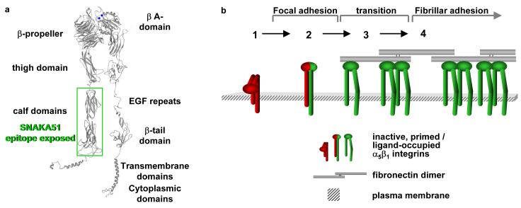
  1. Inactive integrin is diffusely located on the cell surface.

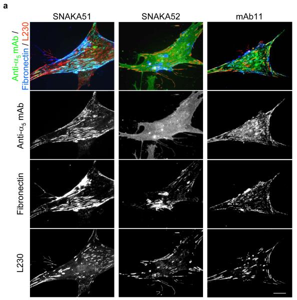
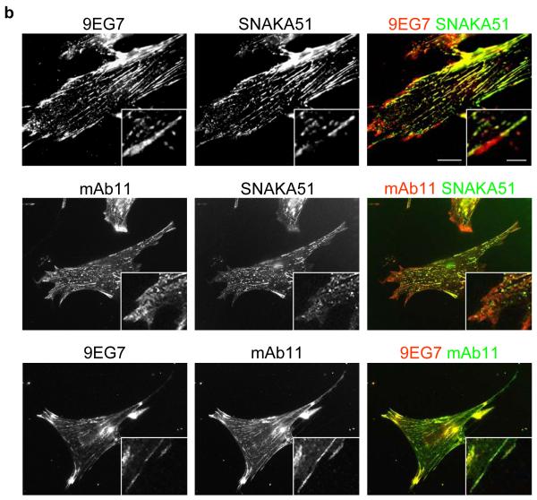
  2. α5β1 located in focal adhesions expresses epitopes reporting a primed β1 conformation (e.g. 9EG7). These integrins may or may not be fully bound by ligand.

  3. Integrin located at the distal edge of focal adhesions has additional SNAKA51 epitope expression. Clustering of this integrin promotes translocation.

  4. Ligated-clustered integrin translocates out of focal adhesions along the actin cytoskeleton, stretching extracellular fibronectin fibrils and driving fibrillogenesis.

References

    1. Akiyama SK, Yamada SS, Chen WT, Yamada KM. Analysis of fibronectin receptor function with monoclonal antibodies: roles in cell adhesion, migration, matrix assembly, and cytoskeletal organization. J Cell Biol. 1989;109:863–75. - PMC - PubMed
    1. Bazzoni G, Shih DT, Buck CA, Hemler ME. Monoclonal antibody 9EG7 defines a novel beta 1 integrin epitope induced by soluble ligand and manganese, but inhibited by calcium. J Biol Chem. 1995;270:25570–7. - PubMed
    1. Beglova N, Blacklow SC, Takagi J, Springer TA. Cysteine-rich module structure reveals a fulcrum for integrin rearrangement upon activation. Nat Struct Biol. 2002;9:282–7. - PubMed
    1. Calderwood DA. Integrin activation. J Cell Sci. 2004;117:657–66. - PubMed
    1. Calvete JJ, Arias J, Alvarez MV, Lopez MM, Henschen A, Gonzalez-Rodriguez J. Further studies on the topography of human platelet glycoprotein IIb. Localization of monoclonal antibody epitopes and the putative glycoprotein IIa- and fibrinogen-binding regions. Biochem J. 1991;273(Pt 3):767–75. - PMC - PubMed

Publication types

MeSH terms